Log in to access this content.
Free for all myUEG account holders. Your access level is set automatically based on your occupation. Medical professionals get full access to all content. If you are a non-medical user, you can only access UEG Week content from congresses you attended.
Not sure what you can access? Learn more about account types.
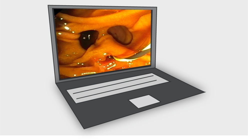
Mistakes in endoscopic retrograde cholangiopancreatography and how to avoid them
Topics
Citation
Published
Log in to access this content.
Free for all myUEG account holders. Your access level is set automatically based on your occupation. Medical professionals get full access to all content. If you are a non-medical user, you can only access UEG Week content from congresses you attended.
Not sure what you can access? Learn more about account types.

Mistakes in cholangioscopy and how to avoid them
Published
Log in to access this content.
Free for all myUEG account holders. Your access level is set automatically based on your occupation. Medical professionals get full access to all content. If you are a non-medical user, you can only access UEG Week content from congresses you attended.
Not sure what you can access? Learn more about account types.
Summary: Autoimmune disease of liver and bile ducts
1 Klinikum rechts der Isar, München, Germany
Event
Topics
Citation
Published
Log in to access this content.
Free for all myUEG account holders. Your access level is set automatically based on your occupation. Medical professionals get full access to all content. If you are a non-medical user, you can only access UEG Week content from congresses you attended.
Not sure what you can access? Learn more about account types.
Best of UEG Week 2024 with Pilar Acedo Nunez and Juozas Kupcinskas on "Bench to Bedside"
Published
Log in to access this content.
Free for all myUEG account holders. Your access level is set automatically based on your occupation. Medical professionals get full access to all content. If you are a non-medical user, you can only access UEG Week content from congresses you attended.
Not sure what you can access? Learn more about account types.
REDUCING RELAPSE RATES IN C. DIFFICILE: IS FIDAXOMICIN THE BETTER OPTION?
1 Norfolk and Norwich University Hospital, Norfolk, United Kingdom
Conference
Topics
Submission format
Citation
Published
Log in to access this content.
Free for all myUEG account holders. Your access level is set automatically based on your occupation. Medical professionals get full access to all content. If you are a non-medical user, you can only access UEG Week content from congresses you attended.
Not sure what you can access? Learn more about account types.
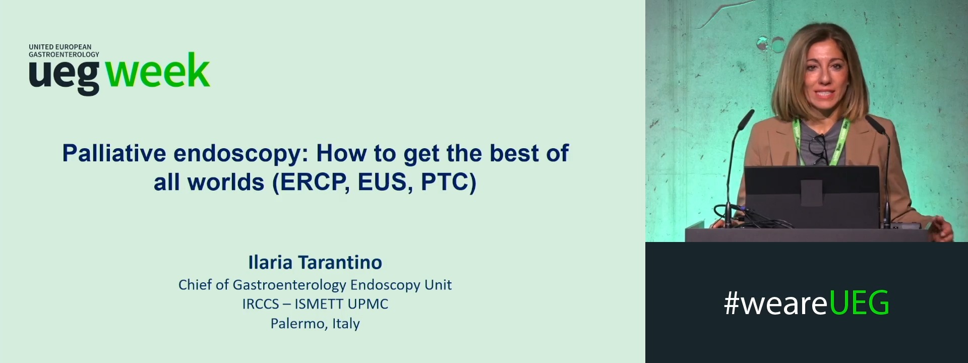
Palliative endoscopy: How to get the best of all worlds (ERCP, EUS, PTC)
1 ISMETT, Palermo, Italy
Event
Citation
Published
Log in to access this content.
Free for all myUEG account holders. Your access level is set automatically based on your occupation. Medical professionals get full access to all content. If you are a non-medical user, you can only access UEG Week content from congresses you attended.
Not sure what you can access? Learn more about account types.
ENDOSCOPIC INTERMUSCULAR DISSECTION FOR EARLY GASTRIC CANCER WITH SEVERE SUBMUCOSAL FIBROSIS
Darshan Parekh 1, Nao Takeuchi 2, Shunya Takayanagi 2, Yoshiaki Kimoto 2, Yohei Minato 2, Hideyuki Chiba 3, Ken Ohata 2
1 Mumbai Institute of Gastroenterology, Mumbai, India
2 NTT Medical Center Tokyo, Tokyo, Japan
3 Omori Red Cross Hospital, Tokyo, Japan
Event
Topics
Submission format
Session