Log in to access this content.
Free for all myUEG account holders. Your access level is set automatically based on your occupation. Medical professionals get full access to all content. If you are a non-medical user, you can only access UEG Week content from congresses you attended.
Not sure what you can access? Learn more about account types.
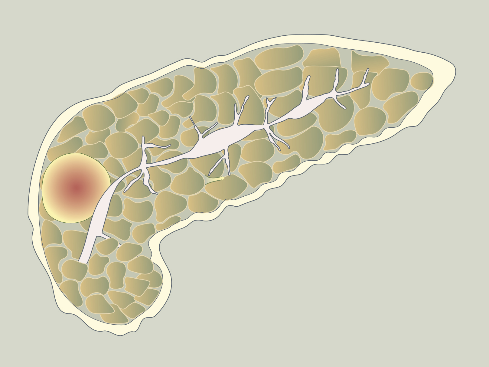
Autoimmune Pancreatitis
Event
Topics
Accreditation status
Duration
Published
Log in to access this content.
Free for all myUEG account holders. Your access level is set automatically based on your occupation. Medical professionals get full access to all content. If you are a non-medical user, you can only access UEG Week content from congresses you attended.
Not sure what you can access? Learn more about account types.

EOSINOPHILIC ESOPHAGITIS AND RISK OF INCIDENT MAJOR ADVERSE CARDIOVASCULAR EVENTS: A NATIONWIDE MATCHED HISTOLOGY COHORT STUDY
Anders Forss 1, Amiko Uchida 2, Bjorn Roelstraete 3, Fahim Ebrahimi 4, John Garber 5, Johan Sundström 6, Jonas F Ludvigsson 7
1 Karolinska Institutet, Stockholm, Sweden|||Karolinska University Hospital, Stockholm, Sweden
2 University of Utah School of Medicine, Salt Lake City, United States|||University of Utah School of Medicine, Salt Lake City, United States
3 Karolinska Institutet, Stockholm, Sweden
4 Karolinska Institutet, Stockholm, Sweden|||Clarunis University Center for Gastrointestinal and Liver Diseases, Basel, Switzerland
5 Harvard Medical School, Boston, United States
6 Uppsala University, Uppsala, Sweden|||University of New South Wales, Sydney, Australia
7 Karolinska Institutet, Stockholm, Sweden|||Örebro University Hospital, Örebro, Sweden|||Columbia University College of Physicians and Surgeons, New York, United States
Conference
Topics
Submission format
Citation
Published
Log in to access this content.
Free for all myUEG account holders. Your access level is set automatically based on your occupation. Medical professionals get full access to all content. If you are a non-medical user, you can only access UEG Week content from congresses you attended.
Not sure what you can access? Learn more about account types.
GASTROINTESTINAL MANIFESTATION AS A PRESENTATION OF HYPEREOSINOPHILIC SYNDROME
Andreia Guimarães 1, Ângela Rodrigues 1, Filomena Barreto 2, Tania Carvalho 1, José Damasceno 1, João Soares 1, Raquel Gonçalves 1
1 Hospital de Braga, Braga, Portugal
2 Laboratório de Anatomia Patológica, Unilabs, Porto, Portugal
Conference
Topics
Submission format
Session
Published
Log in to access this content.
Free for all myUEG account holders. Your access level is set automatically based on your occupation. Medical professionals get full access to all content. If you are a non-medical user, you can only access UEG Week content from congresses you attended.
Not sure what you can access? Learn more about account types.
PANCREATIC CYSTIC LYMPHANGIOMA: A RARE CASE REPORT
1 Military Medical Academy, Cairo, Egypt
2 National Cancer Institute, Cairo University, Cairo, Egypt
Conference
Topics
Submission format
Session
Published
Log in to access this content.
Free for all myUEG account holders. Your access level is set automatically based on your occupation. Medical professionals get full access to all content. If you are a non-medical user, you can only access UEG Week content from congresses you attended.
Not sure what you can access? Learn more about account types.
PREDICTING PANCREATIC CANCER FROM ARTIFICIAL INTELLIGENCE-BASED PANCREATIC VOLUME CHANGES ON CT IMAGING OVER TIME
Jun Nakahodo 1, WATARU UJITA 1, Mizuka Suzuki 1, Masataka Kikuyama 2, Shin-ichiro Horiguchi 1, Masakazu Toi 1, Masanao Kurata 3, Kazurou Chiba 1, Hiroki Tabata 1, Terumi Kamisawa 1, TOSHIRO IIZUKA 1
1 Tokyo Metropolitan Cancer and Infectious Diseases Center, Komagome HospitalH, Tokyo, Japan
2 Tokyo Women's Medical University, Tokyo, Japan
3 University of Tsukuba Mito Clinical Education and Training center, Mito, Japan
Conference
Topics
Submission format
Session
Citation
Published
Log in to access this content.
Free for all myUEG account holders. Your access level is set automatically based on your occupation. Medical professionals get full access to all content. If you are a non-medical user, you can only access UEG Week content from congresses you attended.
Not sure what you can access? Learn more about account types.
WHAT DUODENAL LYMPHANGIECTASIAS HIDE…. INITIAL PRESENTATION OF LUNG CANCER
1 Hospital Universitario Virgen de las Nieves, Granada, Spain
Conference
Topics
Submission format
Session
Published
Log in to access this content.
Free for all myUEG account holders. Your access level is set automatically based on your occupation. Medical professionals get full access to all content. If you are a non-medical user, you can only access UEG Week content from congresses you attended.
Not sure what you can access? Learn more about account types.
ACCURACY OF ENDOSCOPIC ULTRASOUND-GUIDED TISSUE SAMPLING FOR THE CYTO-HISTOLOGICAL DIAGNOSIS OF SOLID PANCREATIC TUMOURS: RESULTS OF A LARGE PROSPECTIVE REGISTRY
YESSICA DOMINGUEZ NOVOA 1, Julio Iglesias-Garcia 1, José Lariño-Noia 1, Daniel De la Iglesia-García 1, Ihab Abdulkader-Nallib 1, Hector Lazare-Iglesias 2, J. Enrique Domínguez Muñoz 1
1 University Hospital of Santiago de Compostela, Santiago de Compostela, Spain|||Research Health Institute of Santiago de Compostela (IDIS), Santiago de Compostela, Spain
2 University Hospital of Santiago de Compostela, Santiago de Compostela, Spain
Conference
Topics