Log in to access this content.
Free for all myUEG account holders. Your access level is set automatically based on your occupation. Medical professionals get full access to all content. If you are a non-medical user, you can only access UEG Week content from congresses you attended.
Not sure what you can access? Learn more about account types.
Mistakes in endoscopic retrograde cholangiopancreatography and how to avoid them
Topics
Citation
Published
Log in to access this content.
Free for all myUEG account holders. Your access level is set automatically based on your occupation. Medical professionals get full access to all content. If you are a non-medical user, you can only access UEG Week content from congresses you attended.
Not sure what you can access? Learn more about account types.

Mistakes in cholangioscopy and how to avoid them
Published
Log in to access this content.
Free for all myUEG account holders. Your access level is set automatically based on your occupation. Medical professionals get full access to all content. If you are a non-medical user, you can only access UEG Week content from congresses you attended.
Not sure what you can access? Learn more about account types.
Episode 9: Harmonising Gastroenterology Training Across Europe
Topics
Published
Log in to access this content.
Free for all myUEG account holders. Your access level is set automatically based on your occupation. Medical professionals get full access to all content. If you are a non-medical user, you can only access UEG Week content from congresses you attended.
Not sure what you can access? Learn more about account types.
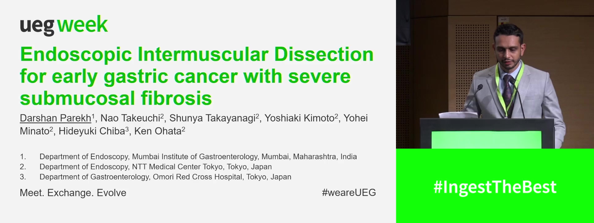
ENDOSCOPIC INTERMUSCULAR DISSECTION FOR EARLY GASTRIC CANCER WITH SEVERE SUBMUCOSAL FIBROSIS
Darshan Parekh 1, Nao Takeuchi 2, Shunya Takayanagi 2, Yoshiaki Kimoto 2, Yohei Minato 2, Hideyuki Chiba 3, Ken Ohata 2
1 Mumbai Institute of Gastroenterology, Mumbai, India
2 NTT Medical Center Tokyo, Tokyo, Japan
3 Omori Red Cross Hospital, Tokyo, Japan
Event
Topics
Submission format
Session
Citation
Published
Log in to access this content.
Free for all myUEG account holders. Your access level is set automatically based on your occupation. Medical professionals get full access to all content. If you are a non-medical user, you can only access UEG Week content from congresses you attended.
Not sure what you can access? Learn more about account types.

EPIDEMIOLOGY OF CHRONIC VIRAL HEPATITIS B/D AND C IN THE VULNERABLE POPULATION IN THE NORTH-EAST AND SOUTH-EAST REGIONS OF ROMANIA – INTERMEDIATE STAGE RESULTS IN THE LIVE(RO)2 - EAST SCREENING
Anca-Victorita Trifan 1, Laura Huiban 2, Cristina Maria Muzica 1, Robert Nastasa 1, Sebastian Zenovia 2, Remus Stafie 1, Ermina Stratina 2, Adrian Rotaru 1, Ana Maria Singeap 1, Camelia Cojocariu 1, Catalin Sfarti 3, Irina Girleanu 2, Stefan Chiriac 1, Horia-Octav Minea 1, Tudor Cuciureanu 1, Carol Stanciu 1
1 “Grigore T. Popa” University of Medicine and Pharmacy, Iasi, Romania|||“St. Spiridon” Hospital, Institute of Gastroenterology and Hepatology, Iasi, Romania
2 “St. Spiridon” Hospital, Institute of Gastroenterology and Hepatology, Iasi, Romania|||“Grigore T. Popa” University of Medicine and Pharmacy, Iasi, Romania
3 “St. Spiridon” Hospital, Institute of Gastroenterology and Hepatology, Iasi, Romania
Conference
Topics
Submission format
Session
Citation
Published
Log in to access this content.
Free for all myUEG account holders. Your access level is set automatically based on your occupation. Medical professionals get full access to all content. If you are a non-medical user, you can only access UEG Week content from congresses you attended.
Not sure what you can access? Learn more about account types.

THE CONCENTRATIONS OF TNFRSF14 AND LIGHT IN THE SERA AND BILE OF THE PATIENTS WITH PRIMARY SCLRELOSING CHOLANGITIS
Sachiko Kanai 1, Hiroaki Fujiwara 2, Suguru Mizuno 3, Takahiro Kishikawa 4, Takuma Nakatsuka 4, Naminatsu Takahara 4, Yousuke Nakai 5, Ryosuke Tateishi 4, Mitsuhiro Fujishiro 4
1 The University of Tokyo, Tokyo, Japan|||The Institute of Medical Science, Asahi Life Foundation, Tokyo, Japan
2 The Institute of Medical Science, Asahi Life Foundation, Tokyo, Japan|||The University of Tokyo, Tokyo, Japan
3 Saitama Medical University, Saitama, Japan
4 The University of Tokyo, Tokyo, Japan
5 The University of Tokyo, Tokyo, Japan|||Tokyo Women's Medical University, Tokyo, Japan
Conference
Topics
Submission format
Session
Citation
Published
Log in to access this content.
Free for all myUEG account holders. Your access level is set automatically based on your occupation. Medical professionals get full access to all content. If you are a non-medical user, you can only access UEG Week content from congresses you attended.
Not sure what you can access? Learn more about account types.

THERE’S STILL A POUCH! REVISIONAL ZENKER PERORAL ENDOSCOPIC MYOTOMY USING A TWO-POINT MYOTOMY TECHNIQUE
Oluwateniola Adeola 1, Benjamin Norton 2, Apostolis Papaefthymiou 2, Andrea Telese 2, Margaret Duku 2, Alexandra Kent 2, Alberto Murino 2, Charlie Murray 2, Gavin Johnson 2, Rehan Haidry 2
1 Queen Mary University London (QMUL), London, United Kingdom
2 Cleveland Clinic London, London, United Kingdom
Conference
Topics