UEG's educational platform for gastroenterology education bringing recordings, courses, guidelines, and more together in one place.
Most popular content
Rate for better recommendations!
Your feedback helps us show you more of what you like and less of what you dislike!
Log in to continue.
This content is part of Gutflix. Log in with your myUEG account, or create one free, to watch it.
Not sure what you can access? Learn more about account types.
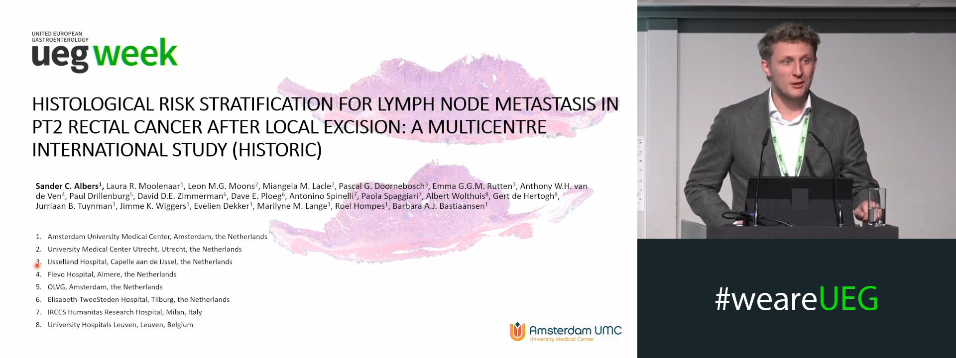
HISTOLOGICAL RISK STRATIFICATION FOR LYMPH NODE METASTASIS IN PT2 RECTAL CANCER AFTER LOCAL EXCISION: A MULTICENTRE INTERNATIONAL STUDY
Sander C. Albers 1, Laura Moolenaar 1, Leon M.G. Moons 2, Miangela M. Lacle 2, Pascal G. Doornebosch 3, Emma G.G.M. Rutten 3, Anthony W.H. van de Ven 4, Paul Drillenburg 5, David D.E. Zimmerman 6, Dave E. Ploeg 6, Antonino Spinelli 7, Paola Spaggiari 7, Albert Wolthuis 8, Gert De Hertogh 8, Jurriaan Tuynman 1, Jimme Wiggers 1, Evelien Dekker 1, Marilyne M. Lange 1, Roel Hompes 1, Barbara A.J. Bastiaansen 1
Affiliations
1 Amsterdam University Medical Center, Amsterdam, Netherlands
2 University Medical Center Utrecht, Utrecht, Netherlands
3 IJsselland Hospital, Capelle aan de IJssel, Netherlands
4 Flevo Hospital, Almere, Netherlands
5 OLVG, Amsterdam, Netherlands
6 Elisabeth-TweeSteden Hospital, Tilburg, Netherlands
7 IRCCS Humanitas Research Hospital, Milan, Italy
8 University Hospital Leuven, Leuven, Belgium

Log in to continue.
This content is part of Gutflix. Log in with your myUEG account, or create one free, to watch it.
Not sure what you can access? Learn more about account types.
I do underwater resection
Affiliations
1 Westmead Hospital Sydney, Westmead, Australia
Log in to continue.
This content is part of Gutflix. Log in with your myUEG account, or create one free, to watch it.
Not sure what you can access? Learn more about account types.
How to define the hot spots?
Affiliations
1 Radboudumc, Nijmegen, Netherlands
Log in to continue.
This content is part of Gutflix. Log in with your myUEG account, or create one free, to watch it.
Not sure what you can access? Learn more about account types.
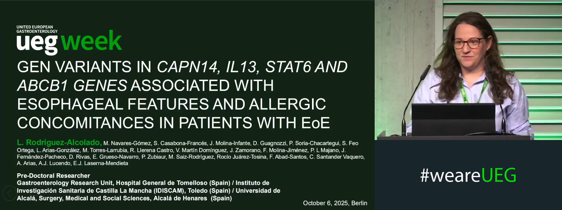
GENE VARIANTS IN CAPN14, IL13, STAT6 AND ABCB1 GENES WERE ASSOCIATED WITH ESOPHAGEAL FEATURES AND ALLERGIC CONCOMITANCES IN PATIENTS WITH EOSINOPHILIC ESOPHAGITIS
Leticia Rodríguez-Alcolado 1, Marcos Navares-Gómez 2, Sergio Casabona-Francés 3, Javier Molina-Infante 4, Danila Guagnozzi 5, Paula Soria-Chacartegui 2, Sara Feo Ortega 6, Laura Arias-González 7, Macarena Torres-Larrubia 8, Ronald Llerena Castro 5, Verónica Martín Domínguez 3, Jose Zamorano 8, Francisca Molina-Jiménez 3, Pedro L Majano 9, Jennifer Fernández-Pacheco 3, Dolores Rivas 8, Elena Grueso-Navarro 7, Pablo Zubiaur 2, Francisco Abad-Santos 10, Cecilio Santander Vaquero 11, Alfredo J. Lucendo 12, Emilio Jose Laserna-Mendieta 7
Affiliations
1 Hospital General de Tomelloso, Gastroenterology Research Unit / Instituto de Investigación Sanitaria de Castilla La Mancha (IDISCAM), Tomelloso, Spain|||Universidad de Alcalá, Alcalá de Henares (Madrid), Spain
2 Hospital Universitario de La Princesa, Clinical Pharmacology / Instituto de Investigación Sanitaria de La Princesa, Madrid, Spain
3 Hospital Universitario de La Princesa, Gastroenterology / Instituto de Investigación Sanitaria de La Princesa, Madrid, Spain
4 Hospital San Pedro de Alcantara, Caceres, Spain|||Centro de Investigación Biomédica en Red Enfermedades Hepáticas y Digestivas (CIBERehd), Madrid, Spain
5 Hospital Universitario Vall d'Hebron, Barcelona, Spain
6 Hospital General de Tomelloso, Pediatrics / Instituto de Investigación Sanitaria de Castilla La Mancha (IDISCAM), Tomelloso, Spain
7 Hospital General de Tomelloso, Gastroenterology Research Unit / Instituto de Investigación Sanitaria de Castilla La Mancha (IDISCAM), Tomelloso, Spain|||Centro de Investigación Biomédica en Red Enfermedades Hepáticas y Digestivas (CIBERehd), Madrid, Spain
8 Hospital San Pedro de Alcantara, Caceres, Spain
9 Hospital Universitario de La Princesa, Gastroenterology / Instituto de Investigación Sanitaria de La Princesa, Madrid, Spain|||Universidad Complutense de Madrid, Madrid, Spain
10 Hospital Universitario de La Princesa, Clinical Pharmacology / Instituto de Investigación Sanitaria de La Princesa, Madrid, Spain|||Centro de Investigación Biomédica en Red Enfermedades Hepáticas y Digestivas (CIBERehd), Madrid, Spain
11 Hospital Universitario de La Princesa, Gastroenterology / Instituto de Investigación Sanitaria de La Princesa, Madrid, Spain|||Centro de Investigación Biomédica en Red Enfermedades Hepáticas y Digestivas (CIBERehd), Madrid, Spain
12 Centro de Investigación Biomédica en Red Enfermedades Hepáticas y Digestivas (CIBERehd), Madrid, Spain|||Hospital General de Tomelloso, Gastroenterology / Instituto de Investigación Sanitaria de Castilla La Mancha (IDISCAM), Tomelloso, Spain
Log in to continue.
This content is part of Gutflix. Log in with your myUEG account, or create one free, to watch it.
Not sure what you can access? Learn more about account types.
Pancreatic lesions? Here’s your cheat sheet for primary care!
Affiliations
1 European Society for Primary Care Gastroenterology (ESPCG), Sant Adrià de Besòs ( Barcelona), Spain
Log in to continue.
This content is part of Gutflix. Log in with your myUEG account, or create one free, to watch it.
Not sure what you can access? Learn more about account types.
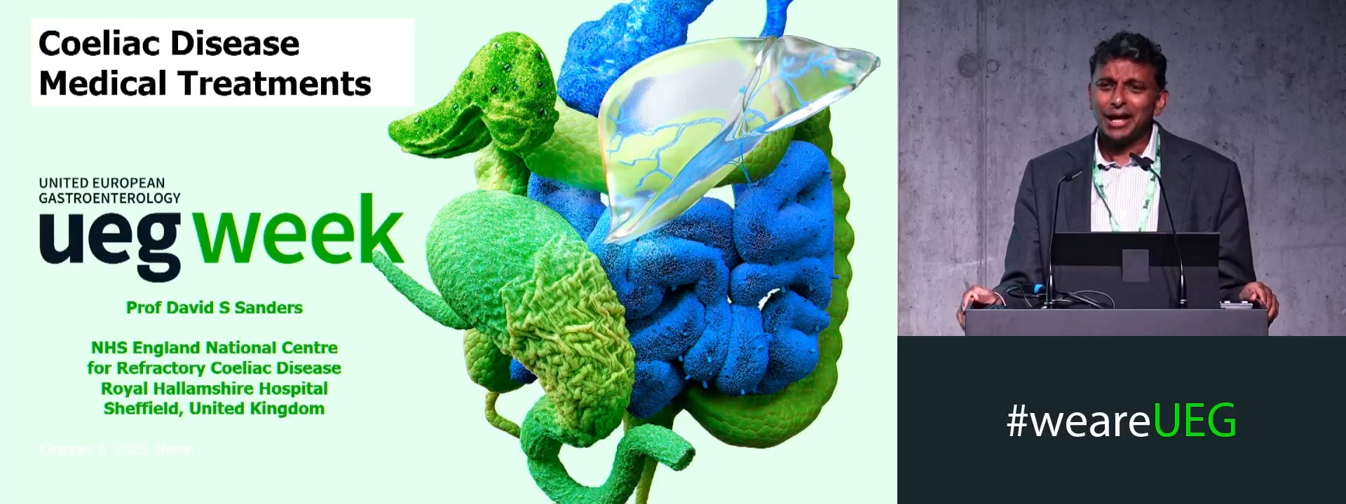
Medical treatment
Affiliations
1 Royal Hallamshire Hospital - NHS Trust, Royal Hallamshire Hospital; Sheffield/GB, Sheffield, United Kingdom
Log in to continue.
This content is part of Gutflix. Log in with your myUEG account, or create one free, to watch it.
Not sure what you can access? Learn more about account types.
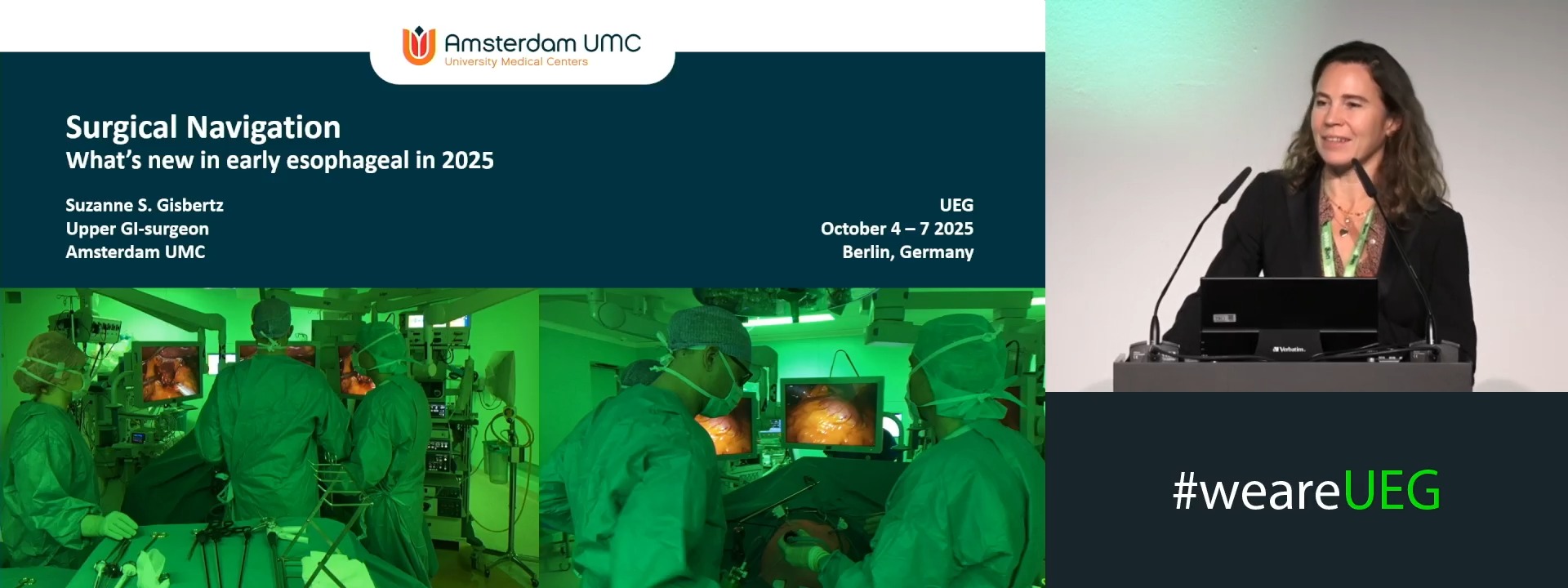
Surgical navigation
Affiliations
1 Amsterdam UMC, Amsterdam, Netherlands