Introduction
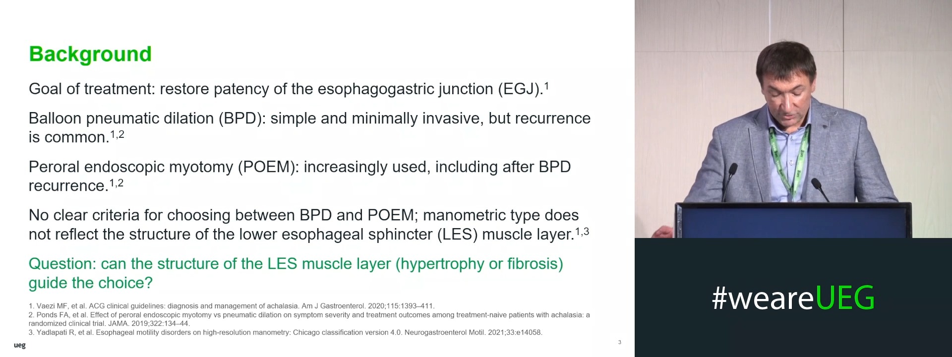
The main goal in treating esophageal achalasia (EA) is to facilitate the patency of the esophagogastric junction (EGJ). Among the current treatment options for EA, pneumatic balloon dilation (BD) is the most common method because it is simple to perform, minimally invasive, and does not require special training. Because recurrence after BD is common, peroral endoscopic myotomy (POEM) has been increasingly adopted. However, clear indications for the use of EA treatment methods have not been definitively determined.
Aims & Methods
The aim of this study was to determine the indications for a differentiated approach to the treatment of EA.
From January 2023 to December 2025, 31 patients diagnosed with EA were evaluated and treated in the digestive system surgery department. High resolution manometry (HRM) classified patients as type I (n=12), type II (n=16), and type III (n=3). The cohort included 12 men (38.7%) and 19 women (61.3%) aged 28 to 74 (40.2 ± 5.4) years. The Eckardt score was used for clinical assessment of EA symptoms. Fluoroscopy was used to determine the diameter of the esophagus, evacuation rate, and length of EGJ narrowing. Endoscopy assessed retained esophageal contents and resistance to passage through the lower esophageal sphincter (LES). EUS with compression elastography was used to determine the structure of the wall of the distal third of the esophagus and LES: a green–yellow pattern indicated hypertrophy, whereas a blue pattern indicated fibrosis. As the first stage of treatment, all patients underwent pneumatic balloon dilation (BD) with balloon systems with a diameter of 35 mm.
Results
After a course of BD (2-5 sessions lasting 60-180 sec) according to the Eckardt score, the symptomatic effectiveness of treatment was 87.1%; the effectiveness of LES relaxation during HRM was 83.8%; the increase in contrast evacuation rate during fluoroscopy was 77.4% after the first course and 80.6% after the repeat BD course. Regardless of the type of EA, the pressure in the LES did not differ (p>0.05), but it decreased significantly (p<0.05) after the BD course. In 2 (6.5%) cases, there were post-dilatation complications: submucosal haematoma and longitudinal tears of the mucous membrane of the distal third of the esophagus. During the follow-up period of up to 16 months, EA recurrence after the first course of BD was observed in 22.6% of cases: type I – 2 (6.5%), type II – 4 (12.9%), type III – 1 (3.2%). In 19.4% of cases of EA recurrence, repeated courses of BD were ineffective. To determine the causes of recurrence in these patients, EUS was performed and fibrosis with thickening of the wall of the distal third of the esophagus – 3.26 (3.12; 3.38) mm and LES – 5.65 (5.14; 6.14) mm was diagnosed. The presence of fibrosis contributed to post-dilatation complications and EA recurrence. In 19.4% of cases of EA recurrence following BD, POEM was performed. According to the Eckardt score, symptomatic treatment efficacy and increased contrast evacuation rate during fluoroscopy at follow-up periods up to 12 months were observed in 5 out of 6 patients.
Conclusion
EA recurrence is associated with fibrotic transformation of the wall of the distal third of the esophagus and LES. The use of HRM allows assessing the effectiveness of BD. EUS enables differentiation between hypertrophy and fibrotic transformation of the distal esophageal wall and the LES; the latter may warrant myotomy.