UEG's content platform for gastroenterology education bringing recordings, courses, guidelines, and more together in one place.
Trending on Gutflix
Rate for better recommendations!
Your feedback helps us show you more of what you like and less of what you dislike!
Log in to continue.
This content is part of Gutflix. Log in with your myUEG account, or create one free, to watch it.
Not sure what you can access? Learn more about account types.
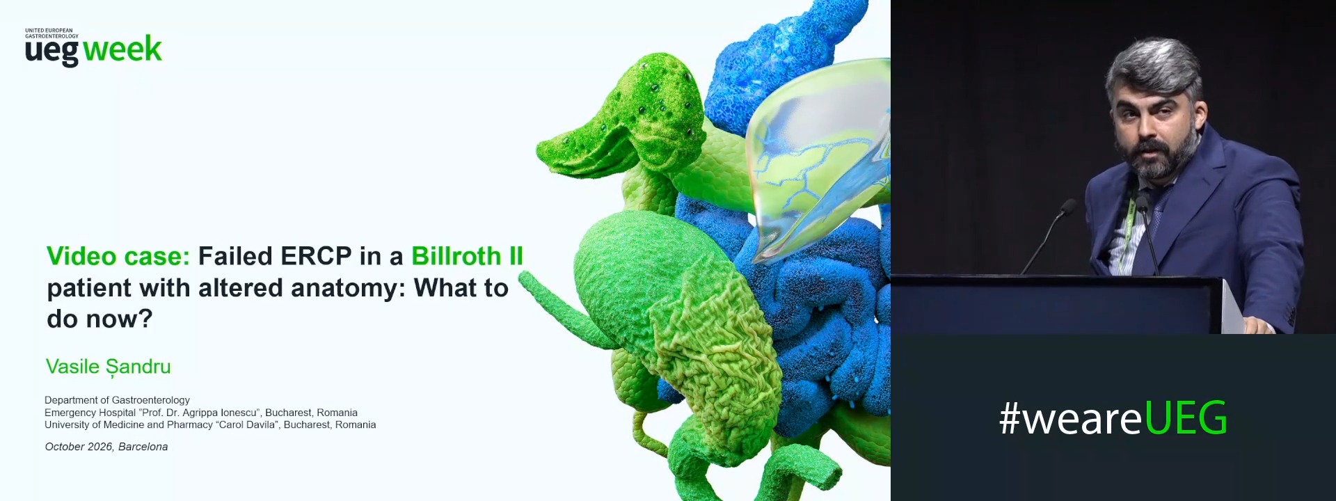
Video case: Failed ERCP in a Billroth II patient with altered anatomy: What to do now?
Affiliations
1 Bucharest Emergency Hospital, Bucharest, Romania
Log in to continue.
This content is part of Gutflix. Log in with your myUEG account, or create one free, to watch it.
Not sure what you can access? Learn more about account types.
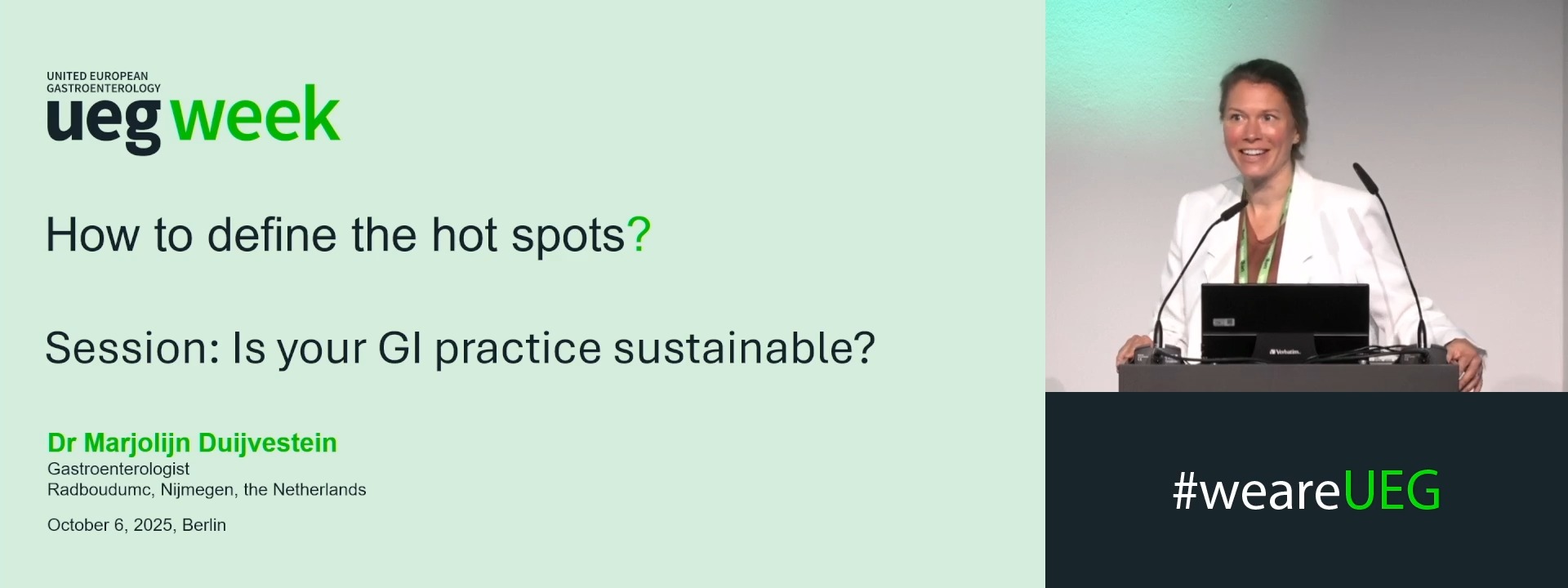
How to define the hot spots?
Affiliations
1 Radboudumc, Nijmegen, Netherlands

Log in to continue.
This content is part of Gutflix. Log in with your myUEG account, or create one free, to watch it.
Not sure what you can access? Learn more about account types.
NEUTROPHIL-FIBROBLAST CROSSTALK THROUGH JAK/STAT SIGNALING AND IL-8 AS A NEW MECHANISM OF CROHN’S DISEASE IMMUNOFIBROSIS
Efstratios Gavriilidis 1, Anastasia Maria Natsi 1, Christina Antoniadou 1, Dionysios Kogias 1, Victoria Tsironidou 1, NIKOS KAFALIS 1, Stylianos Didaskalou 2, Maria Koffa 2, Alexandra Giatromanolaki 1, Georgios Kouklakis 1, Konstantinos Ritis 1, Panagiotis Skendros 1
Affiliations
1 Democritus University of Thrace, Faculty of Medicine, Alexandroupolis, Greece
2 Democritus University of Thrace, Department of Molecular Biology and Genetics, Alexandroupolis, Greece

Log in to continue.
This content is part of Gutflix. Log in with your myUEG account, or create one free, to watch it.
Not sure what you can access? Learn more about account types.
T1b squamous cancer (Complete Session)

Log in to continue.
This content is part of Gutflix. Log in with your myUEG account, or create one free, to watch it.
Not sure what you can access? Learn more about account types.
How to create AI for Barrett's oesophagus and how to integrate it in its management
Affiliations
1 Academic Medical Centre - Gastroenterology & Hepatology, Academic Medical Centre; Amsterdam/NL, Amsterdam, Netherlands
2 Eindhoven University of Technology, Eindhoven, Netherlands