UEG's content platform for gastroenterology education bringing recordings, courses, guidelines, and more together in one place.
Trending on Gutflix
Rate for better recommendations!
Your feedback helps us show you more of what you like and less of what you dislike!
Log in to continue.
This content is part of Gutflix. Log in with your myUEG account, or create one free, to watch it.
Not sure what you can access? Learn more about account types.
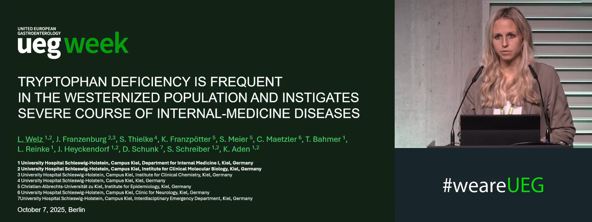
TRYPTOPHAN DEFICIENCY IS FREQUENT IN THE WESTERNIZED POPULATION AND INSTIGATES SEVERE COURSE OF INTERNAL-MEDICINE DISEASES
Lina Welz 1, Jeanette Franzenburg 1, Sascha Thielke 1, Katrin Franzpötter 2, Sarah Meier 2, Corina Maetzler 1, Olaf Schneider 3, Jan Heyckendorf 1, Thomas Bahmer 1, Domagoj Schunk 1, Stefan Schreiber 4,1, Konrad Aden 1
Affiliations
1 University Hospital Schleswig-Holstein, Campus Kiel, Kiel, Germany
2 Christian-Albrechts-Universität zu Kiel, Kiel, Germany
3 Cerascreen, Leipzig, Germany
4 University Hospital Schleswig-Holstein, Kiel, Germany
Log in to continue.
This content is part of Gutflix. Log in with your myUEG account, or create one free, to watch it.
Not sure what you can access? Learn more about account types.
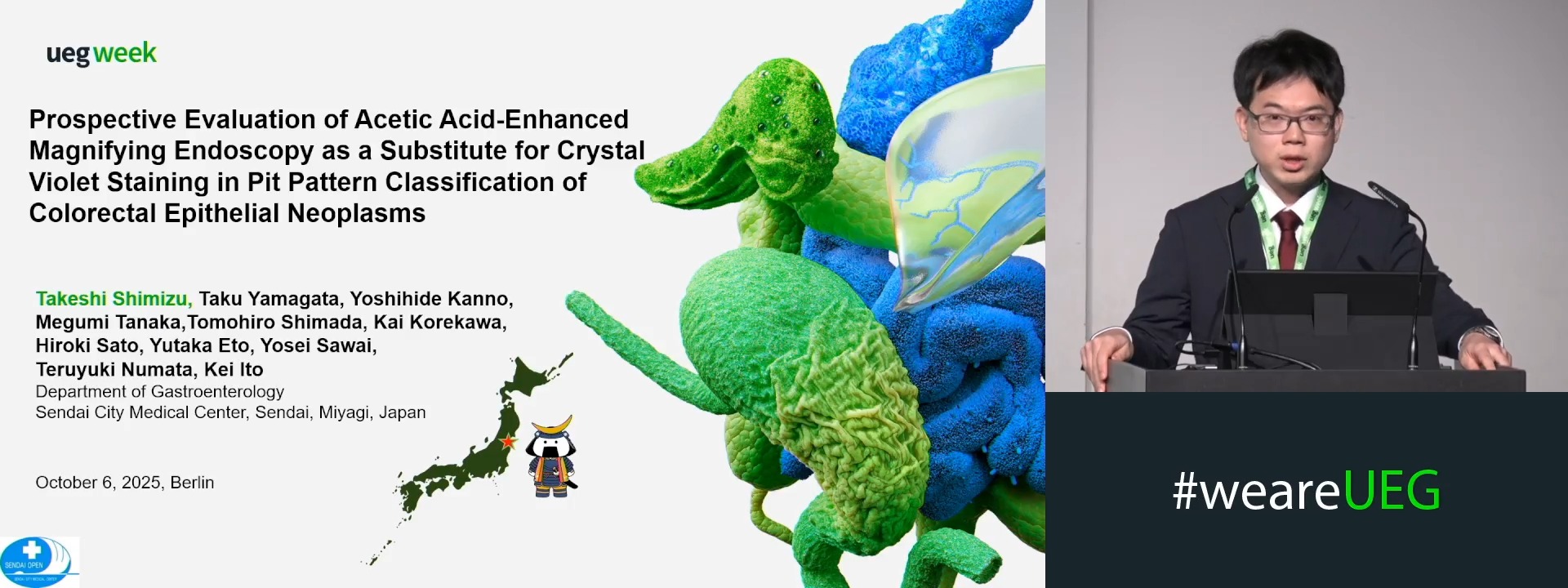
PROSPECTIVE EVALUATION OF ACETIC ACID-ENHANCED MAGNIFYING ENDOSCOPY AS A SUBSTITUTE FOR CRYSTAL VIOLET STAINING IN PIT PATTERN CLASSIFICATION OF COLORECTAL EPITHELIAL NEOPLASMS
Takeshi Shimizu 1, Taku Yamagata 1, Yoshihide Kanno 1, Megumi Tanaka 1, Tomohiro Shimada 1, Kai Korekawa 1, Hiroki Sato 1, Yutaka Eto 1, Yosei Sawai 1, Teruyuki Numata 1, Kei Ito 1
Affiliations
1 Sendai City Medical Center, Sendai, Japan
Log in to continue.
This content is part of Gutflix. Log in with your myUEG account, or create one free, to watch it.
Not sure what you can access? Learn more about account types.
Specific key function of gut microbiota
Affiliations
1 Deutsches Zentrum für Infektionsforschung, Braunschweig, Germany

Log in to continue.
This content is part of Gutflix. Log in with your myUEG account, or create one free, to watch it.
Not sure what you can access? Learn more about account types.
How to improve quality in colonoscopy screening: Is AI the answer?
Affiliations
1 University of Oslo / Showa University, Oslo, Norway
Log in to continue.
This content is part of Gutflix. Log in with your myUEG account, or create one free, to watch it.
Not sure what you can access? Learn more about account types.
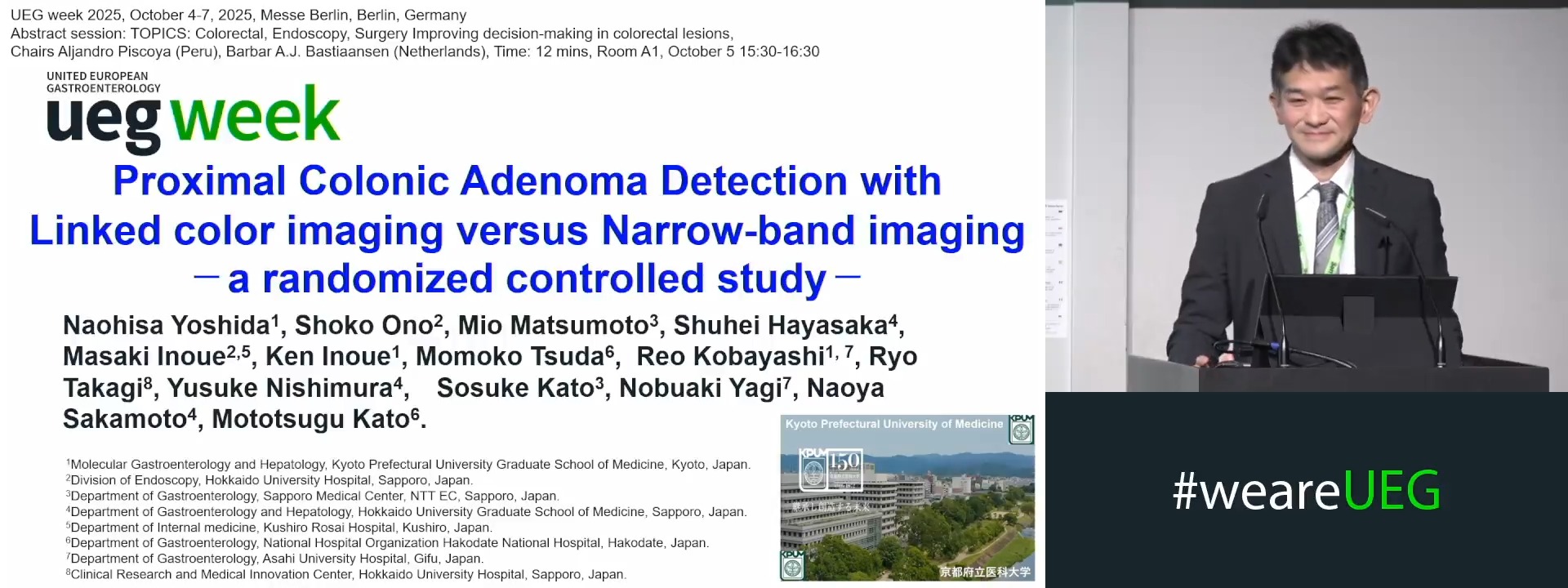
PROXIMAL COLONIC ADENOMA DETECTION WITH LINKED COLOR IMAGING VERSUS NARROW-BAND IMAGING-A RANDOMIZED CONTROLLED STUDY
Naohisa Yoshida 1, SHOKO ONO 2, Mio Matsumoto 3, Shuhei Hayasaka 4, Masaki Inoue 2, Ken Inoue 1, Momoko Tsuda 5, Reo Kobayashi 1, Ryo Takagi 2, Yusuke Nishimura 6, Sosuke Kato 3, Nobuaki Yagi 7, Naoya Sakamoto 8, Mototsugu Kato 5
Affiliations
1 Kyoto Prefectural University of Medicine, Kyoto, Japan
2 Hokkaido University Hospital, Sapporo, Japan
3 Sapporo Medical Center, NTT EC, Sapporo, Sapporo, Japan
4 Hokkaido University Graduate School of Medicine, Sapporo, Japan
5 National Hospital Organization Hakodate National Hospital, Hakodate, Japan
6 Kushiro Rosai Hospital, Kushiro, Japan
7 Asahi University Hospital, Gifu, Japan
8 Hokkaido University Graduate School of Medicine, Hokkaido, Japan