UEG's content platform for gastroenterology education bringing recordings, courses, guidelines, and more together in one place.
Trending on Gutflix
Rate for better recommendations!
Your feedback helps us show you more of what you like and less of what you dislike!
Log in to continue.
This content is part of Gutflix. Log in with your myUEG account, or create one free, to watch it.
Not sure what you can access? Learn more about account types.
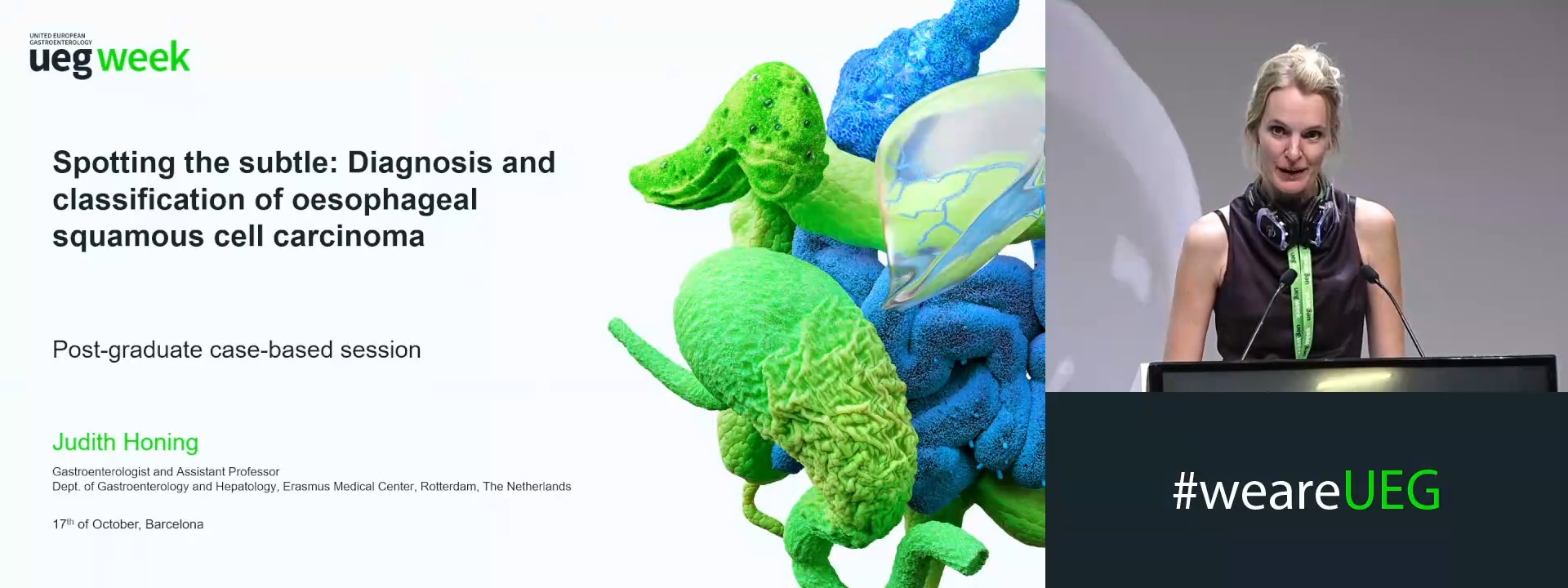
Spotting the subtle: Diagnosis and classification of early squamous cell carcinoma
Affiliations
1 Erasmus Medical Center Rotterdam, Rotterdam, Netherlands
Log in to continue.
This content is part of Gutflix. Log in with your myUEG account, or create one free, to watch it.
Not sure what you can access? Learn more about account types.
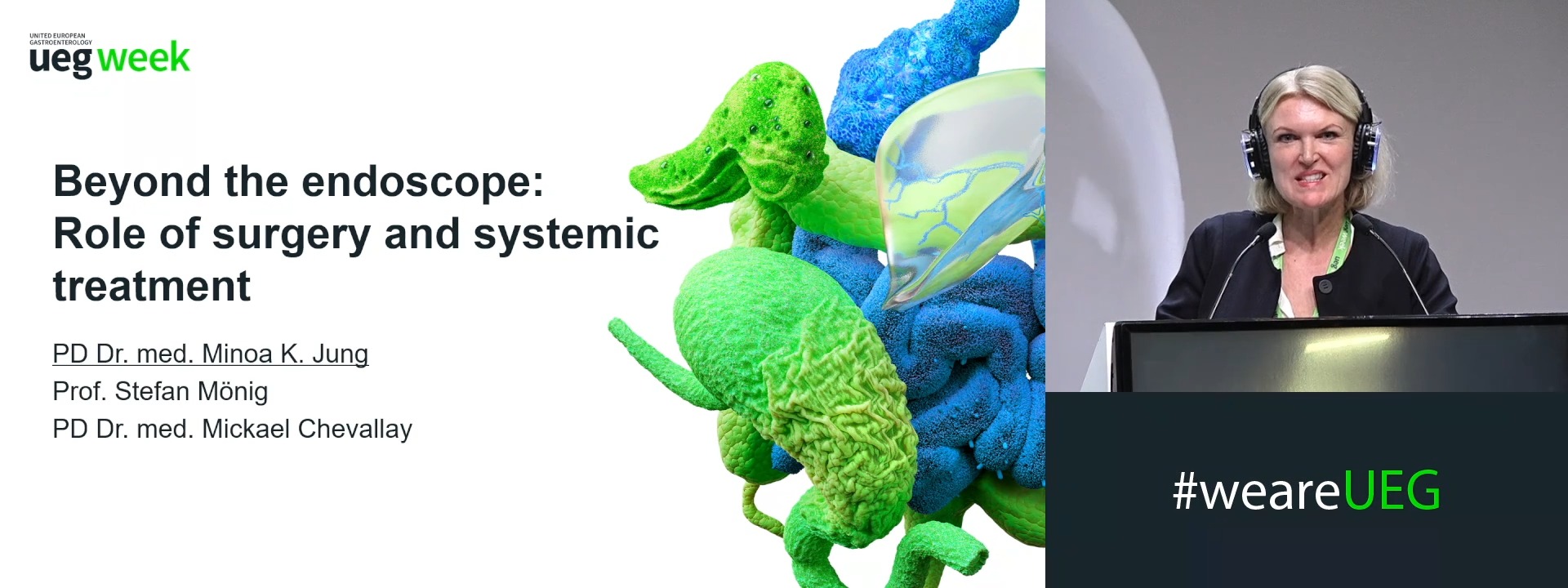
Beyond the endoscope: Role of surgery and systemic treatment
Affiliations
1 University Hospitals of Geneva, Geneva, Switzerland
Log in to continue.
This content is part of Gutflix. Log in with your myUEG account, or create one free, to watch it.
Not sure what you can access? Learn more about account types.
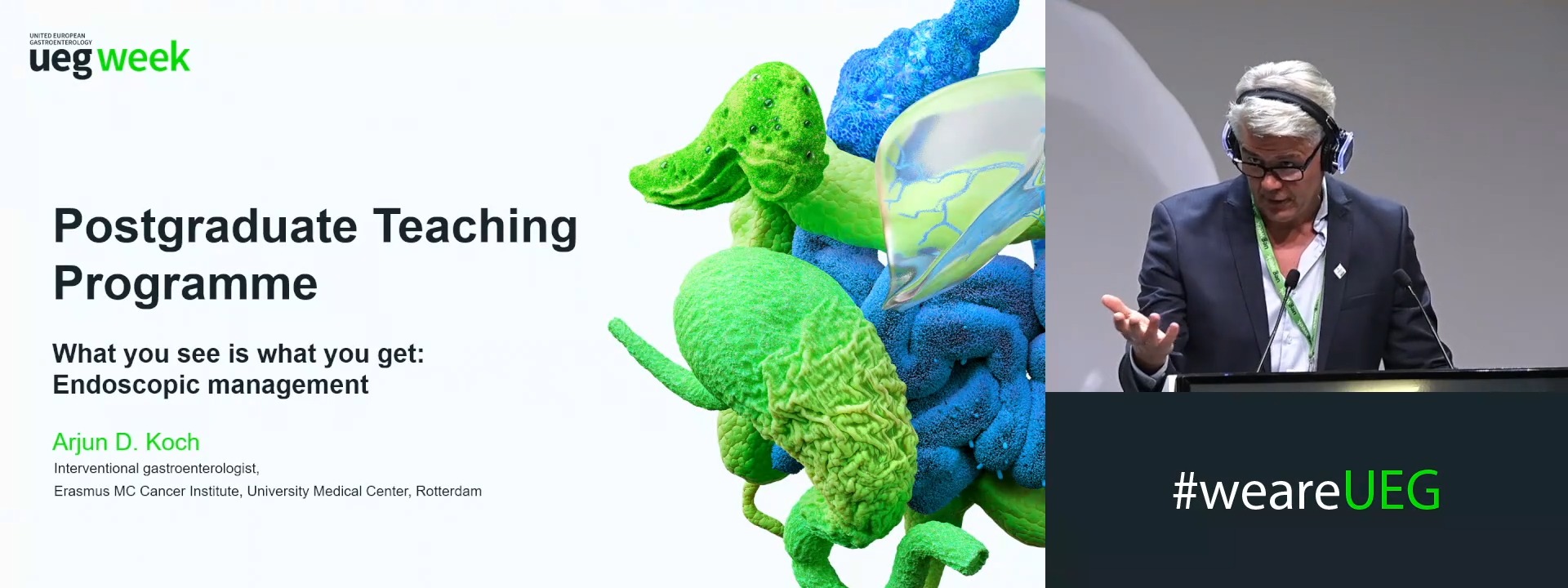
What you see is what you get: Endoscopic management
Affiliations
1 Erasmus MC - University Medical center, Rotterdam, Netherlands
Log in to continue.
This content is part of Gutflix. Log in with your myUEG account, or create one free, to watch it.
Not sure what you can access? Learn more about account types.
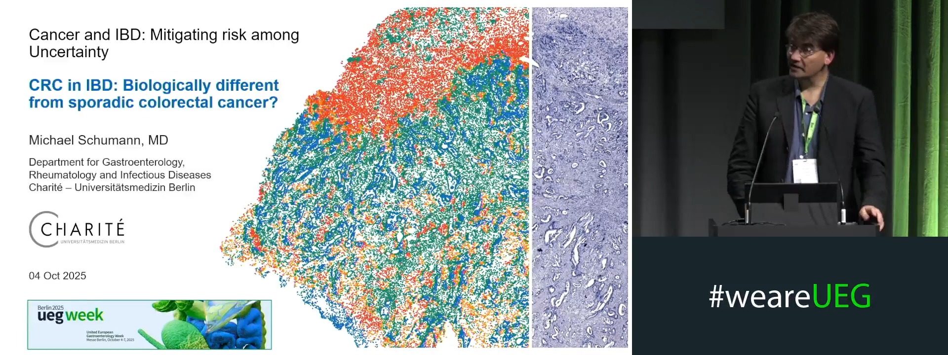
CRC in IBD: Biologically different from sporadic colorectal cancer?
Affiliations
1 Charité Berlin, Berlin, Germany

Log in to continue.
This content is part of Gutflix. Log in with your myUEG account, or create one free, to watch it.
Not sure what you can access? Learn more about account types.
T1b squamous cancer (Complete Session)
Log in to continue.
This content is part of Gutflix. Log in with your myUEG account, or create one free, to watch it.
Not sure what you can access? Learn more about account types.
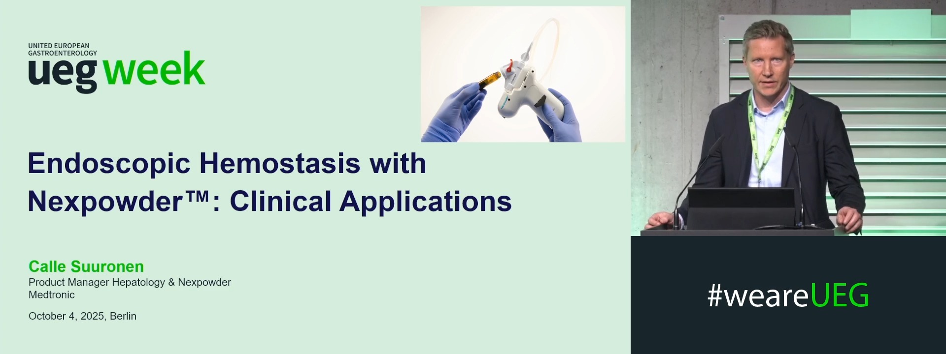
Endoscopic hemostasis with Nexpowder™* : Clinical applications
Affiliations
1 Medtronic AB, Kista, Sweden