UEG's content platform for gastroenterology education bringing recordings, courses, guidelines, and more together in one place.
Trending on Gutflix
Rate for better recommendations!
Your feedback helps us show you more of what you like and less of what you dislike!
Log in to continue.
This content is part of Gutflix. Log in with your myUEG account, or create one free, to watch it.
Not sure what you can access? Learn more about account types.
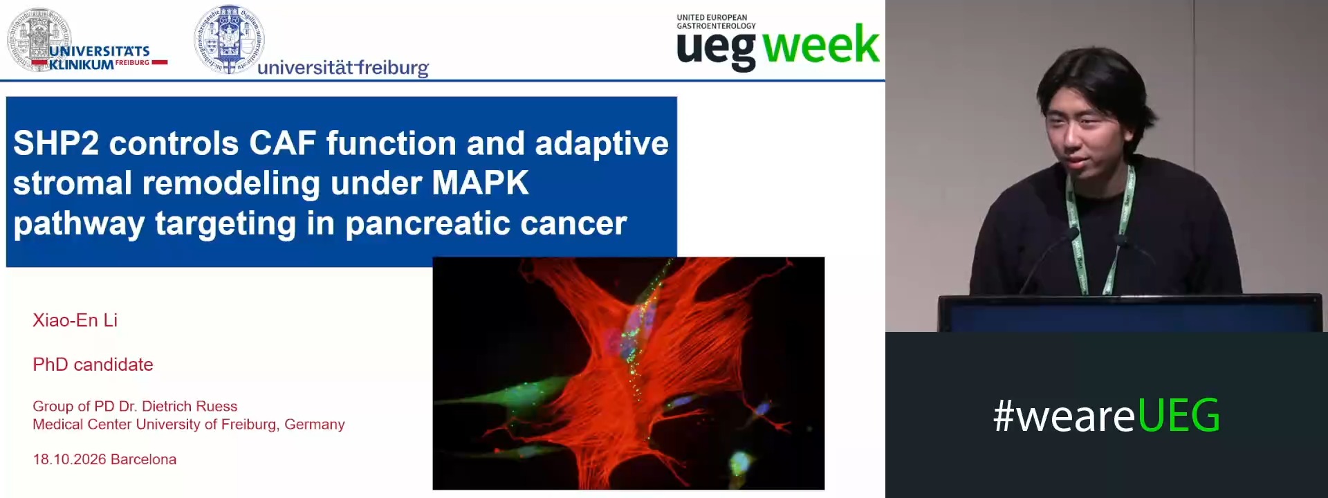
SHP2 REGULATES CAF ACTIVITY AND ADAPTIVE STROMAL REMODELING DURING VERTICAL RAS PATHWAY INHIBITION IN PANCREATIC CANCER
Steffen Keller 1, Xun Chen 1, Tonmoy Das 2, Xiao-En Li 1, Hanna Scheffold 1, Philipp Hafner 1, Asma Alrawashdeh 1, Huda Jumaa 1, Dominik von Elverfeldt 1, Wilfried Reichardt 1, Geoffroy Andrieux 2, Melanie Boerries 2,3, Stefan Fichtner-Feigl 1, Dietrich Alexander Ruess 1,3
Affiliations
1 University Medical Center Freiburg, Freiburg, Germany
2 Institute of Medical Bioinformatics and Systems Medicine, University of Freiburg, Freiburg, Germany
3 German Cancer Consortium (DKTK), Freiburg, Germany
Log in to continue.
This content is part of Gutflix. Log in with your myUEG account, or create one free, to watch it.
Not sure what you can access? Learn more about account types.
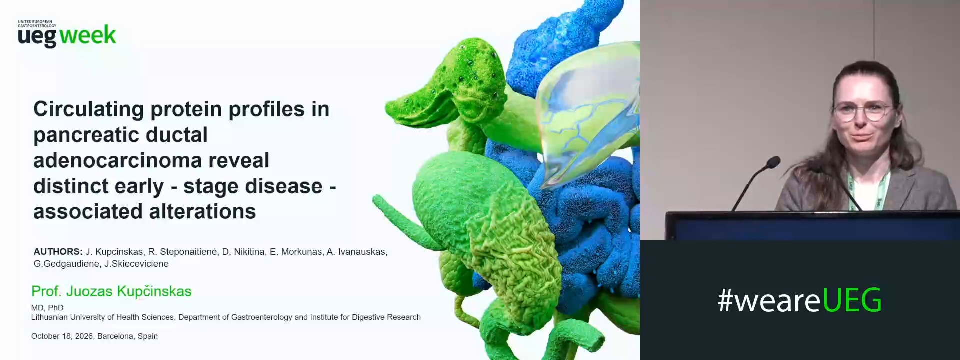
CIRCULATING PROTEIN PROFILES IN PANCREATIC DUCTAL ADENOCARCINOMA REVEAL DISTINCT EARLY-STAGE DISEASE-ASSOCIATED ALTERATIONS
Juozas Kupcinskas 1,2, Rūta Steponaitienė 2, Darja Nikitina 2, Egidijus Morkunas 1, Audrius Ivanauskas 1, Greta Gedgaudiene 2, Jurgita Skieceviciene 2
Affiliations
1 Lithuanian University of Health Sciences, Kaunas, Lithuania
2 Lithuanian University Of Health Sciences, Kaunas, Lithuania