UEG's educational platform for gastroenterology education bringing recordings, courses, guidelines, and more together in one place.
Most popular content
Rate for better recommendations!
Your feedback helps us show you more of what you like and less of what you dislike!
Log in to access this content.
Free for all myUEG account holders. Your access level is set automatically based on your occupation. Medical professionals get full access to all content. If you are a non-medical user, you can only access UEG Week content from congresses you attended.
Not sure what you can access? Learn more about account types.

FUNCTIONAL VERIFICATION AND MECHANISM OF FAM129C GENE MUTATION IN ACHALASIA
1 Zhongshan Hospital, Shanghai, China
Conference
Topics
Submission format
Citation
Published
Log in to access this content.
Free for all myUEG account holders. Your access level is set automatically based on your occupation. Medical professionals get full access to all content. If you are a non-medical user, you can only access UEG Week content from congresses you attended.
Not sure what you can access? Learn more about account types.
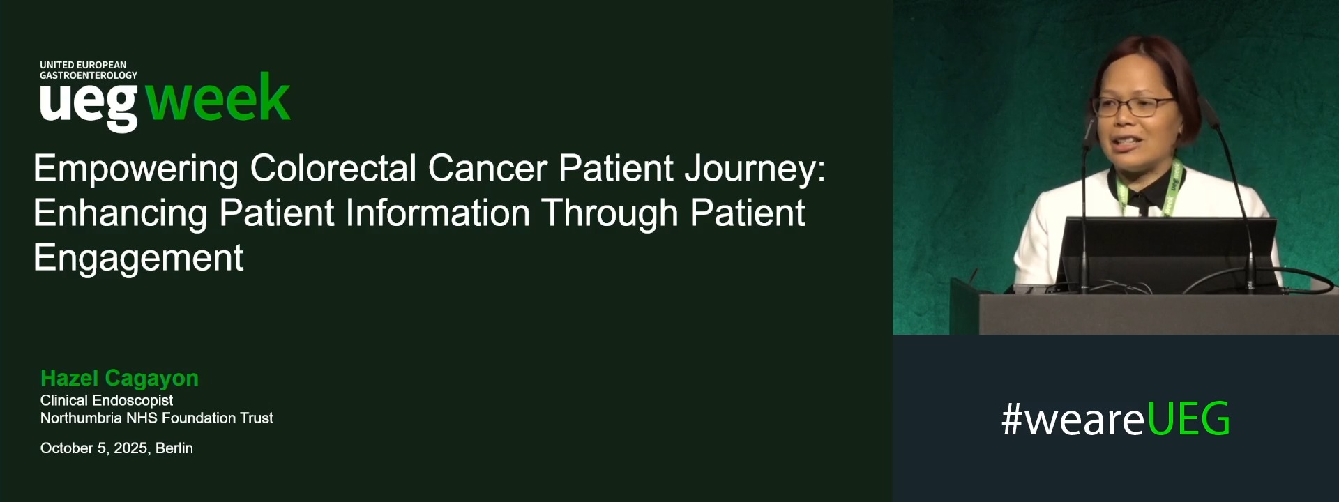
EMPOWERING COLORECTAL CANCER PATIENT JOURNEY: ENHANCING PATIENT INFORMATION THROUGH PATIENT ENGAGEMENT
1 Northumbria NHS Trust, Northumberland, United Kingdom
Event
Topics
Submission format
Citation
Published
Log in to access this content.
Free for all myUEG account holders. Your access level is set automatically based on your occupation. Medical professionals get full access to all content. If you are a non-medical user, you can only access UEG Week content from congresses you attended.
Not sure what you can access? Learn more about account types.
THE EVALUATION OF ENDOSCOPISTS’ SKILL IMPROVEMENT THROUGH D-LECS WITH ESD USING THE ESD ASSESSMENT SCORE
Hiroyuki Aoyagi 1, Yukino Shinazawa 1, Kaisei Iwamoto 1, Hayato Fujita 1, Shyunichi Ueda 1, Taiga Saitou 1, Chie Hattori 1, Kouhei Yasuda 1, Norihiko Ogawa 1, Tsuyoshi Suda 1, Yoshihide Naito 1, Kenkei Hasatani 1, Yasuharu Kaizaki 1, Toshiyuki Okuda 1, Tamon Miyanaga 1, Itasu Ninomiya 1
1 Fukui Prefectural Hospital, Fukui, Japan
Conference
Topics
Submission format
Citation
Published
Log in to access this content.
Free for all myUEG account holders. Your access level is set automatically based on your occupation. Medical professionals get full access to all content. If you are a non-medical user, you can only access UEG Week content from congresses you attended.
Not sure what you can access? Learn more about account types.
LIVER SINOSOIDAL ENDOTHELIAL CELL CONTRIBUTES TO LEUKOCYTE RECRUITMENT IN LIVER FIBROSIS THROUGH A CYCLOOXYGENASE-2-DEPENDENT MECHANISM
Yang Tai 1, Yang Xiao 1, Xin Quan 1, Linhao Zhang 1, Huan Tong 1, bo wei 1, Hao Wu 1, Jinhang Gao 2, Chengwei Tang 1
1 West China Hospital, Sichuan University, Chengdu, China
2 West China Hospital, Sichuan University, Chengdu, China|||West China Hospital, Sichuan University, Chengdu, China
Conference
Topics
Submission format
Citation
Published
Log in to access this content.
Free for all myUEG account holders. Your access level is set automatically based on your occupation. Medical professionals get full access to all content. If you are a non-medical user, you can only access UEG Week content from congresses you attended.
Not sure what you can access? Learn more about account types.
THE EFFICACY OF BILIARY COVERED METAL STENT OVER CONVENTIONAL TREATMENT IN ENDOSCOPIC RETROGRADE CHOLANGIOPANCREATOGRAPHY RELATED TYPE II RETROPERITONEAL PERFORATION
1 Kyungpook National University School of Medicine, Daegu, Korea, Republic of
Conference
Topics
Submission format
Session
Citation
Published
Log in to access this content.
Free for all myUEG account holders. Your access level is set automatically based on your occupation. Medical professionals get full access to all content. If you are a non-medical user, you can only access UEG Week content from congresses you attended.
Not sure what you can access? Learn more about account types.
EXPLORING BIOMARKERS OF SYSTEMIC OXIDATIVE STRESS AND PLACENTAL INSUFFICIENCY IN PREGNANT WOMEN WITH INFLAMMATORY BOWEL DISEASES
Hannah Holstein 1, Dianne Gelien Bouwknegt 1, Marijn C. Visschedijk 1, Marian L. C. Bulthuis 1, Marjan Reinders-Luinge 1, Mirthe H. Schoots 1, Harry van Goor 1, Sanne J. Gordijn 1, Gerard Dijkstra 1, Arno R. Bourgonje 2
1 University of Groningen, University Medical Center Groningen, Groningen, Netherlands
2 University of Groningen, University Medical Center Groningen, Groningen, Netherlands|||Icahn School of Medicine at Mount Sinai, New York, United States
Conference
Topics
Submission format
Session
Citation
Published
Log in to access this content.
Free for all myUEG account holders. Your access level is set automatically based on your occupation. Medical professionals get full access to all content. If you are a non-medical user, you can only access UEG Week content from congresses you attended.
Not sure what you can access? Learn more about account types.
IBD HEALTH EQUITY SUMMIT: A CALL TO ACTION
1 Pfizer Inc, New York, United States
Conference
Topics
Submission format
Session