Introduction
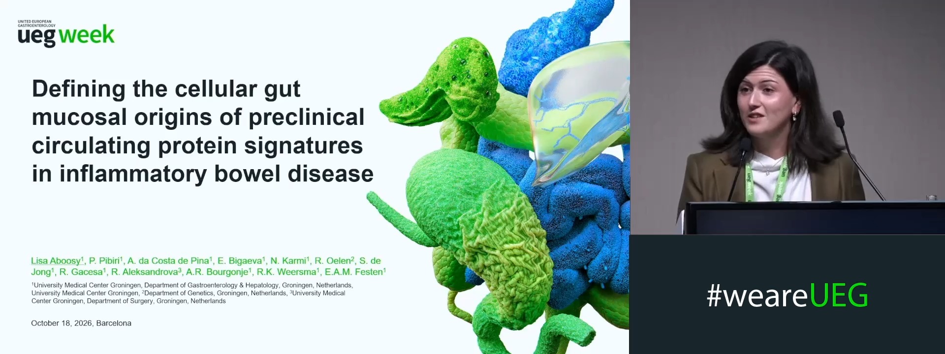
The research paradigm in inflammatory bowel disease (IBD) is shifting toward biomarker-based prediction and early detection before clinical onset. Circulating protein signatures can identify individuals at risk of IBD years before diagnosis, supporting the existence of a preclinical phase1. However, the cellular origin of these proteins remains unclear, limiting interpretation of whether they reflect early disease biology, subclinical mucosal inflammation or other processes.
Aims & Methods
To investigate the cellular origin of preclinical circulating protein signatures in IBD, genes encoding a selected set of previously reported Crohn’s disease (CD) and ulcerative colitis (UC) circulating protein signatures were mapped onto in-house UMCG single-cell RNA-sequencing datasets from CD, UC, and healthy controls (HC)1. CD proteins included CXCL9, IL6, MMP10, CCL20, MDK, CXCL17, DNER, GPNMB, and CX3CL1, while UC proteins included STAMBP, SIRT2, SCAMP3, ADAM8, GZMB, ABL1, CCL11, CD5, TNFRSF6B, MMP10, CDKN1A, and CXCL9. CD data were derived from terminal ileum (159 biopsies; 151 patients; 96 female; mean age 46 years), while UC data were derived from colon (78 biopsies; 53 patients; 28 female; mean age 41 years). Colonic biopsies from HC (n=5) served as controls. Cells were annotated into 51 (CD) and 54 (UC/HC) cell types using published references2-4. For each protein-encoding gene, cell types with highest average expression were identified and differential expression between inflamed and non-inflamed tissue was assessed using the Wilcoxon rank-sum test with Benjamini-Hochberg correction (adjusted P<0.05).
Results
Across IBD, genes encoding preclinical proteins were predominantly expressed in intestinal mucosal immune cells, whereas in HC expression was mainly localized to stromal subsets. In CD, highest average expression was observed in Th17 tissue-resident memory cells, activated dendritic cells (DC), and inflammatory and resident macrophages. Six of nine CD-associated biomarkers (CXCL9, IL6, DNER, MDK, GPNMB, MMP10) showed significant differential expression between inflamed vs. non-inflamed tissue, with strongest associations for IL6 in inflammatory macrophages (adjusted P (P)=2.15x10⁻¹⁴), MMP10 in transitional stromal 3(C3+) cells (P=1.93x10⁻⁵) and CXCL9 in activated DCs (P=1.03x10⁻⁴). In UC, highest average expression was likewise observed across immune subsets, including DCs, cycling T- and B-cells, inflammatory monocytes, natural killer cells (NK), CD8+IL17+ T-cells, plasmacytoïd DCs, and macrophages. Nine of twelve UC-associated biomarkers (STAMBP, SCAMP3, ADAM8, GZMB, ABL1, CCL11, CD5, CDKN1A, CXCL9) showed significant inflammation‑associated differences, with strongest associations for CCL11 in WNT2B+Fos-lo1 fibroblasts (P=9.87x10-47), CDKN1A in inflammatory monocytes (P=3.50x10⁻¹¹) and GZMB in NKs (P=1.39x10⁻⁸). In HCs, highest average expression was mainly observed in stromal subsets including RSPO3+ and WNT2B+Fos-lo1 fibroblasts, endothelial, microvascular and glial cells. TNFRSF6B remained undetectable across all gut mucosal subsets.
Conclusion
Transcripts from previously identified circulating proteins in CD and UC are predominantly expressed by gut mucosal immune cells. Several show inflammation-associated expression differences, suggesting these proteins reflect local mucosal inflammatory activity in established IBD. Transcripts without such differences may arise from alternative cell states or tissue not captured in gut mucosa. These findings provide biological context for preclinical IBD protein signatures and help interpret the processes they may represent.
References
1. Grännö, O. et al. (2025) Gastroenterology. https://doi.org/10.1053/j.gastro.2024.11.006
2. Smillie, C. S. et al. (2019) Cell. https://doi.org/10.1016/j.cell.2019.06.029
3. Martin, J. C. et al. (2019) Cell. https://doi.org/10.1016/j.cell.2019.08.008
4. Elmentaite, R. et al. (2020) Cell. https://doi.org/10.1016/j.devcel.2020.11.010
Disclosure
Sample processing was performed by Takeda.