UEG's educational platform for gastroenterology education bringing recordings, courses, guidelines, and more together in one place.
Trending on Gutflix
Rate for better recommendations!
Your feedback helps us show you more of what you like and less of what you dislike!
Log in to continue.
This content is part of Gutflix. Log in with your myUEG account, or create one free, to watch it.
Not sure what you can access? Learn more about account types.
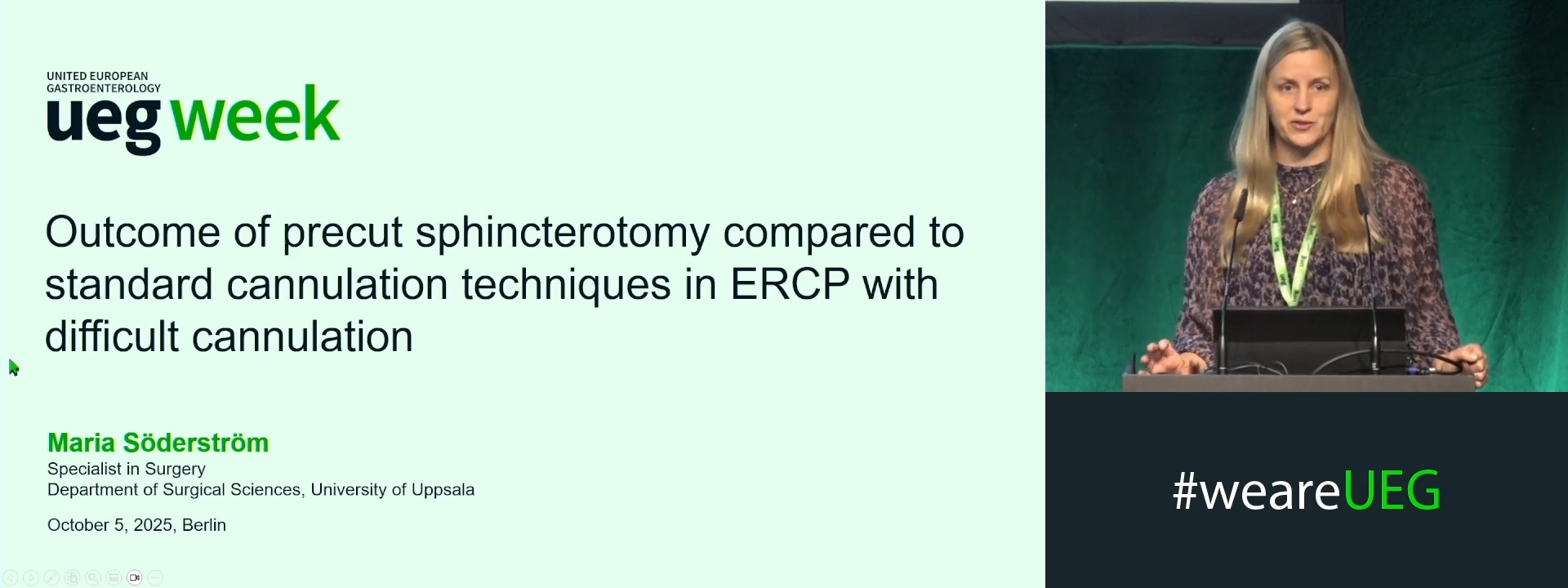
OUTCOME OF PRECUT SPHINCTEROTOMY COMPARED TO STANDARD CANNULATION TECHNIQUES IN ERCP WITH DIFFICULT CANNULATION
Affiliations
1 Uppsala University, Uppsala, Sweden
2 Karolinska Institutet, Stockholm, Sweden
Log in to continue.
This content is part of Gutflix. Log in with your myUEG account, or create one free, to watch it.
Not sure what you can access? Learn more about account types.
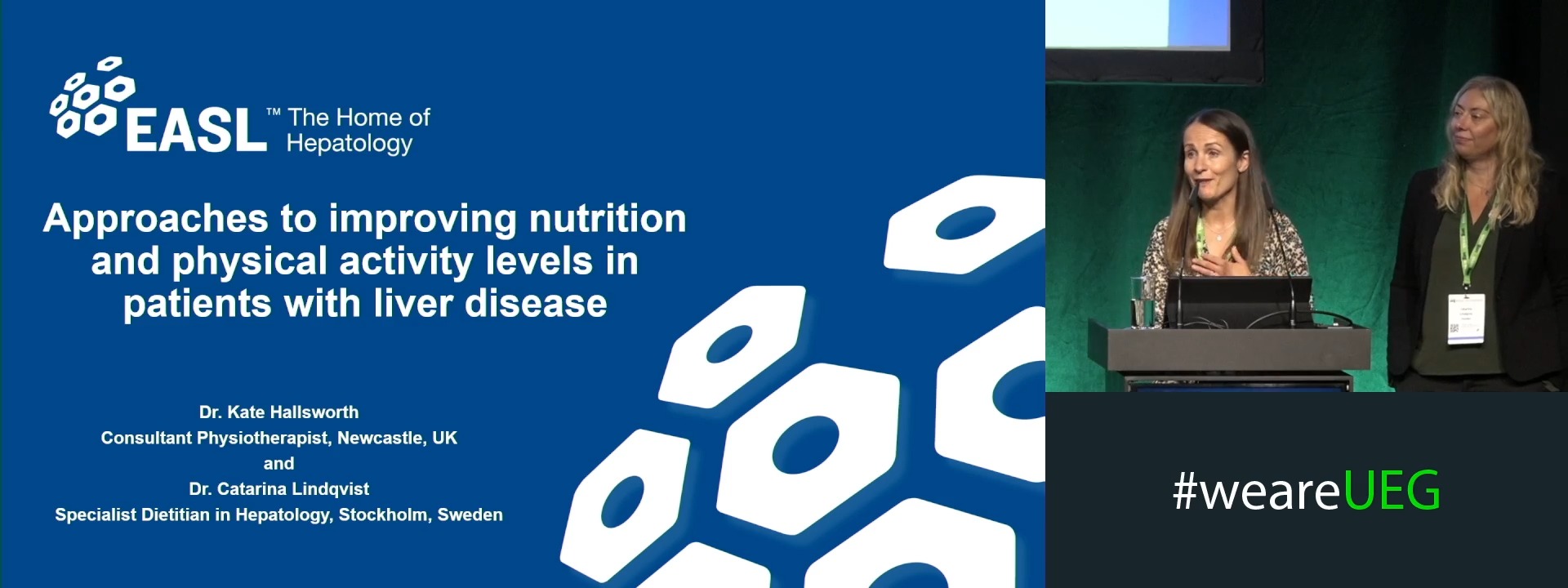
Approaches to improve nutrition and physical activity levels in patients with liver disease
Affiliations
1 NIHR Newcastle Biomedical Research Centre, Newcastle upon Tyne, United Kingdom
2 None, None, Sweden
Log in to continue.
This content is part of Gutflix. Log in with your myUEG account, or create one free, to watch it.
Not sure what you can access? Learn more about account types.
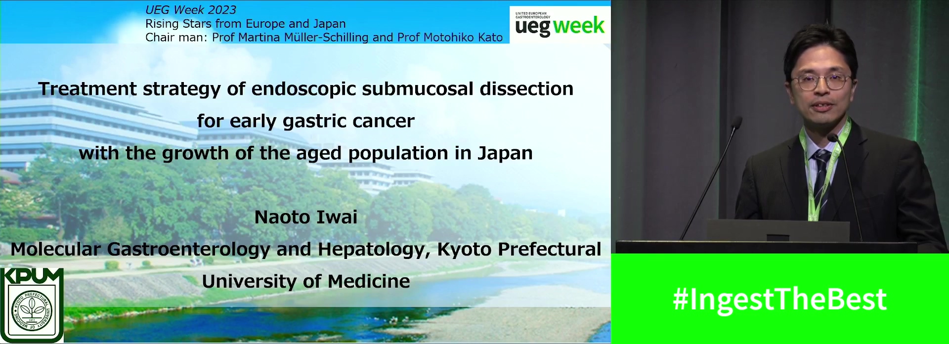
Treatment strategy of endoscopic submucosal dissection for early gastric cancer with the growth of the aged population in Japan
Affiliations
1 , Kyoto Prefectural University of Medicine Graduate School of Medical Scien, Kyoto, Japan
Log in to continue.
This content is part of Gutflix. Log in with your myUEG account, or create one free, to watch it.
Not sure what you can access? Learn more about account types.
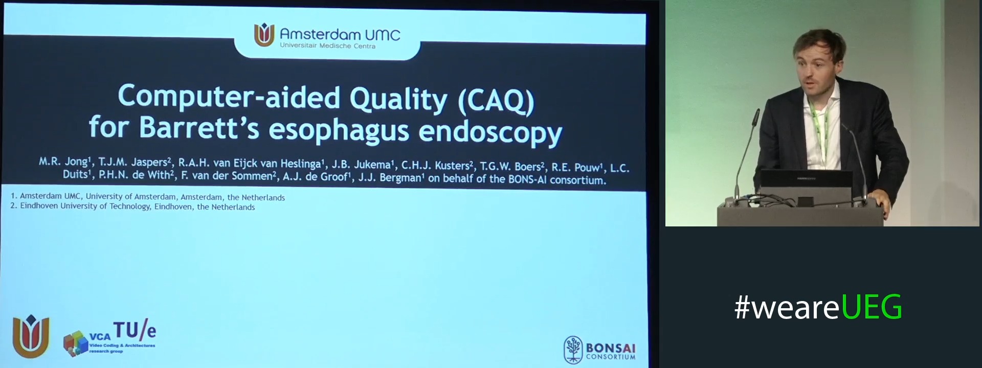
COMPUTER AIDED QUALITY CONTROL SYSTEM IMPROVES BARRETT’S NEOPLASIA DETECTION IN AN EX-VIVO SETTING
Martijn R. Jong 1, Tim Jaspers 2, Rixta van Eijck van Heslinga 1, Jelmer Jukema 1, Carolus Kusters 2, Tim Boers 2, Roos E. Pouw 3, Lucas Duits 1, Peter H. N. de With 2, Fons van der Sommen 2, Jeroen de Groof 1, Jacques J. Bergman 1
Affiliations
1 Amsterdam UMC, Amsterdam, Netherlands
2 TU Eindhoven, Eindhoven, Netherlands
3 UMC Utrecht, Utrecht, Netherlands