UEG's content platform for gastroenterology education bringing recordings, courses, guidelines, and more together in one place.
Trending on Gutflix
Rate for better recommendations!
Your feedback helps us show you more of what you like and less of what you dislike!
Log in to continue.
This content is part of Gutflix. Log in with your myUEG account, or create one free, to watch it.
Not sure what you can access? Learn more about account types.
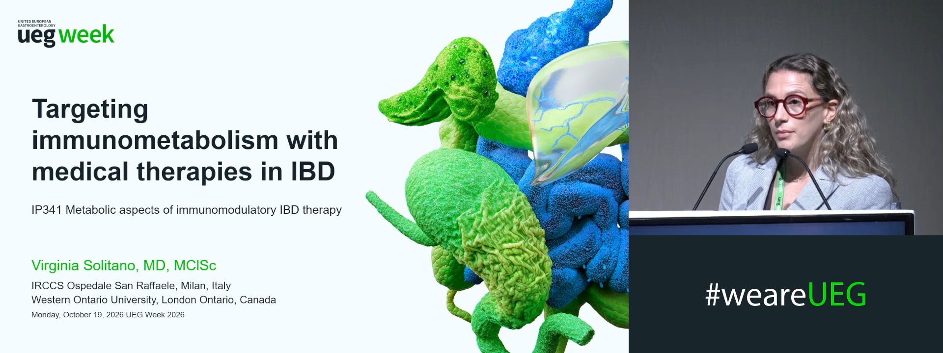
Targeting immunometabolism with medical therapies in IBD
Affiliations
1 IRCCS Ospedale San Raffaele, Milan, Italy
Log in to continue.
This content is part of Gutflix. Log in with your myUEG account, or create one free, to watch it.
Not sure what you can access? Learn more about account types.
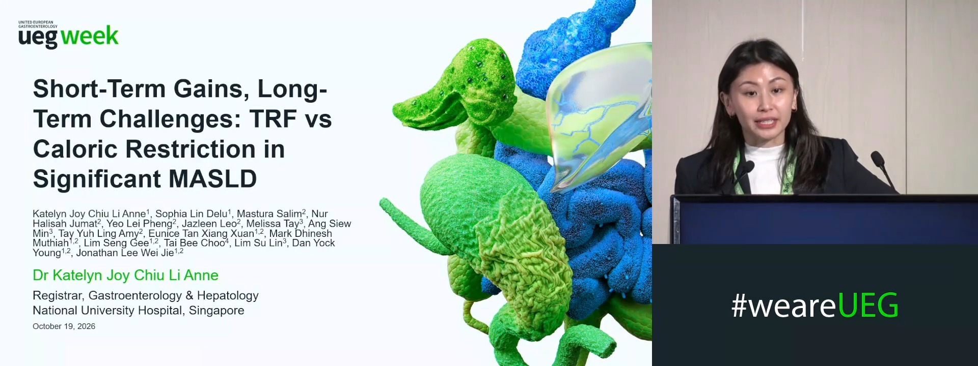
SHORT-TERM GAINS, LONG-TERM CHALLENGES: A PILOT RANDOMIZED CONTROLLED TRIAL FOR TIME-RESTRICTED FEEDING VERSUS CALORIC RESTRICTION IN SIGNIFICANT MASLD
Katelyn Chiu 1, Sophia Lin Delu 1, Jonathan Lee 2, Mastura Binte Salim 3, Nur Halisah Jumat 3, Lei Pheng Yeo 3, Jazleen Leo 3, Melissa Tay 4, Siew Min Ang 4, Amy Tay Yuh Ling 3, Eunice Tan 1, Mark Muthiah 1, Seng Gee Lim 1, Bee Choo Tai 3, Su Lin Lim 4, Dan Yock Young 1
Affiliations
1 National University Hospital, Singapore, Singapore, Singapore
2 National University Health System, Singapore, Singapore, Singapore
3 National University of Singapore, Singapore, Singapore
4 National University Hospital, Singapore, Singapore
Log in to continue.
This content is part of Gutflix. Log in with your myUEG account, or create one free, to watch it.
Not sure what you can access? Learn more about account types.
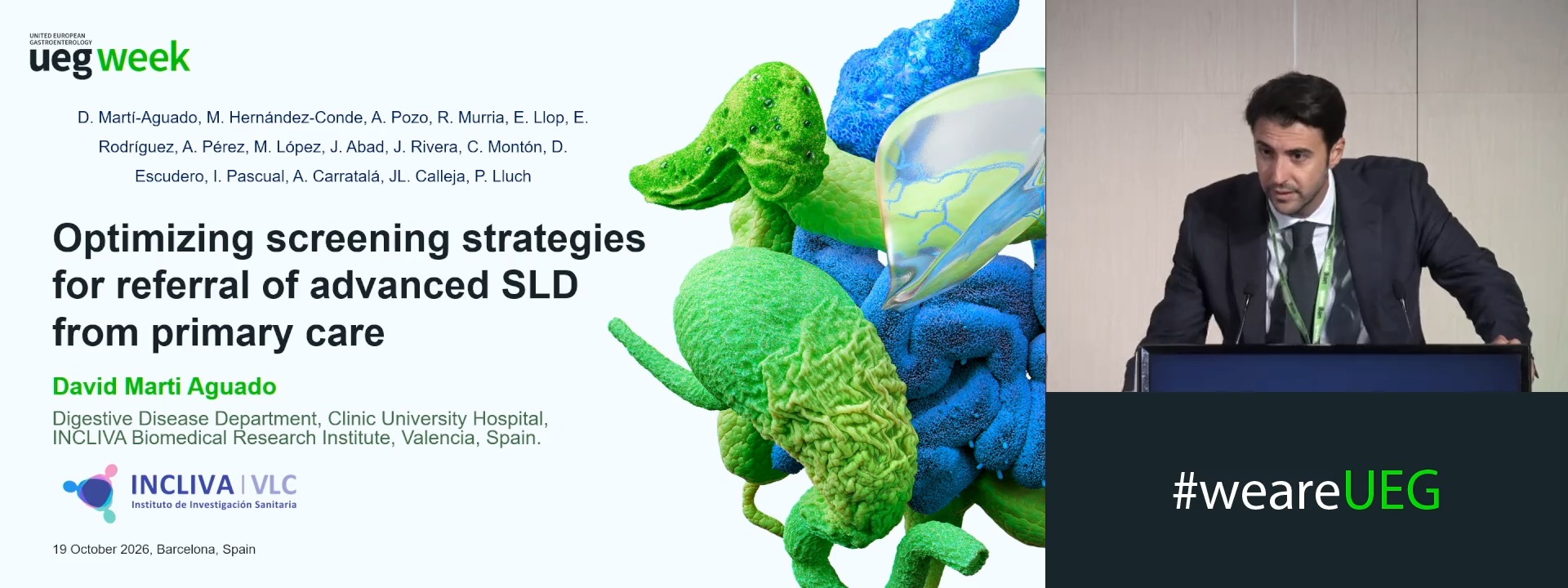
OPTIMIZING SCREENING STRATEGIES FOR REFERRAL OF ADVANCED STEATOTIC LIVER DISEASE FROM PRIMARY CARE
David Marti Aguado 1, Marta Hernandez Conde 2, Adela Pozo Giraldez 1, Rosa Murria 1, Enrique Rodríguez-Borja 1, Ana Perez 2, Elba Llop 3, Marta Lopez Gomez 4, Javier Abad 4, Jesus Manuel Rivera Esteban 2, Cristina Monton 1, Desamparados Escudero-Garcia 5, Isabel Pascual-Moreno 6, Arturo Carratalá-Calvo 1, Jose Luis Calleja 4, Paloma Lluch García 1
Affiliations
1 Clinic University Hospital of Valencia, INCLIVA Health Research Institute, Valencia, Spain
2 Puerta de Hierro University Hospital, Madrid, Spain
3 Gastroenterology, Hospital Puerta de Hierro, Madrid, Spain, Madrid, Spain
4 Hospital Universitario Puerta de Hierro, Pozuelo De Alarcon, Spain
5 University of Valencia, Valencia, Spain
6 Hospital Clinico Universitario, Valencia, Spain
Log in to continue.
This content is part of Gutflix. Log in with your myUEG account, or create one free, to watch it.
Not sure what you can access? Learn more about account types.
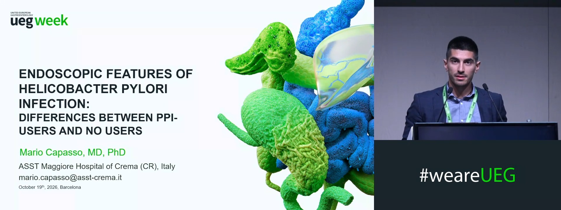
ENDOSCOPIC FEATURES OF HELICOBACTER PYLORI INFECTION: DIFFERENCES BETWEEN PPI-USERS AND NO USERS
Mario Capasso 1, Michael Visentin 1, Samanta Romeo 1, Roberto Bertè 1, Flavio Metelli 1, Antonio Cerrone 1, Stefania Bertolazzi 1, Simona Albano 1, Saverio Alicante 1, Claudio Londoni 1, Guido Manfredi 1
Affiliations
1 ASST MAggiore Hospital Cema, Crema (CR), Italy
Log in to continue.
This content is part of Gutflix. Log in with your myUEG account, or create one free, to watch it.
Not sure what you can access? Learn more about account types.
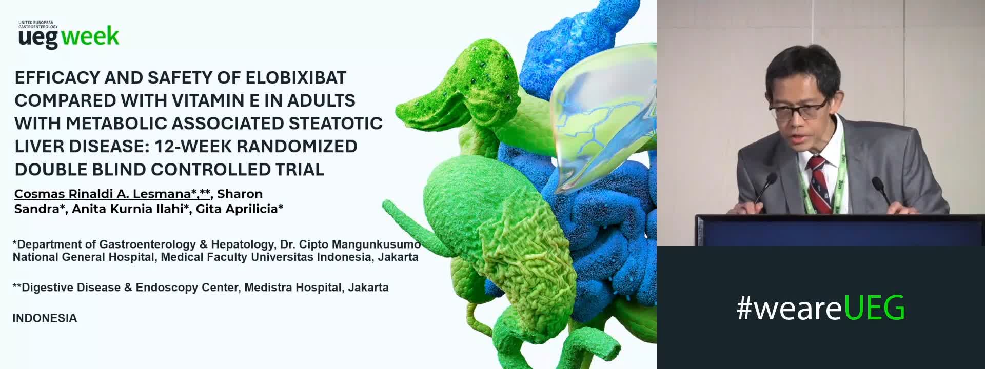
EFFICACY AND SAFETY OF ELOBIXIBAT COMPARED WITH VITAMIN E IN ADULTS WITH METABOLIC ASSOCIATED STEATOTIC LIVER DISEASE: A 12-WEEK RANDOMIZED DOUBLE BLIND CONTROLLED TRIAL
Cosmas Rinaldi A Lesmana 1,2, Sharon Sandra 3, Anita Kurnia Ilahi 3, Gita Aprilicia 3, Irsan Hasan 1
Affiliations
1 Dr. Cipto Mangunkusumo National General Hospital, Medical Faculty Universitas Indonesia, Jakarta, Indonesia
2 Medistra Hospital, Jakarta, Indonesia
3 Dr. Cipto Mangunkusumo National General Hospital, Jakarta, Indonesia