UEG's content platform for gastroenterology education bringing recordings, courses, guidelines, and more together in one place.
Trending on Gutflix
Rate for better recommendations!
Your feedback helps us show you more of what you like and less of what you dislike!

Log in to continue.
This content is part of Gutflix. Log in with your myUEG account, or create one free, to watch it.
Not sure what you can access? Learn more about account types.
When is the time to call the surgeon?
Affiliations
1 University of North Carolina / NCRBSI, London, United Kingdom
Log in to continue.
This content is part of Gutflix. Log in with your myUEG account, or create one free, to watch it.
Not sure what you can access? Learn more about account types.
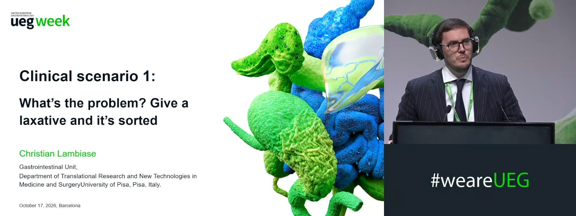
Clinical scenario 1: What’s the problem? Give a laxative and it’s sorted
Affiliations
1 Gastrointestinal Unit, Department of Translational Sciences and New Technologies in Medicine and Surgery, University of Pisa, Pisa, Italy
Log in to continue.
This content is part of Gutflix. Log in with your myUEG account, or create one free, to watch it.
Not sure what you can access? Learn more about account types.
Introduction: Chronic constipation: How to diagnose and treat this frequent condition in clinical practice
Affiliations
1 University of Nottingham, Nottingham, United Kingdom

Log in to continue.
This content is part of Gutflix. Log in with your myUEG account, or create one free, to watch it.
Not sure what you can access? Learn more about account types.
Summary: Chronic constipation: How to diagnose and treat this frequent condition in clinical practice
Affiliations
1 University Of Sydney, Sydney, Australia