UEG's content platform for gastroenterology education bringing recordings, courses, guidelines, and more together in one place.
Trending on Gutflix
Rate for better recommendations!
Your feedback helps us show you more of what you like and less of what you dislike!
Log in to continue.
This content is part of Gutflix. Log in with your myUEG account, or create one free, to watch it.
Not sure what you can access? Learn more about account types.
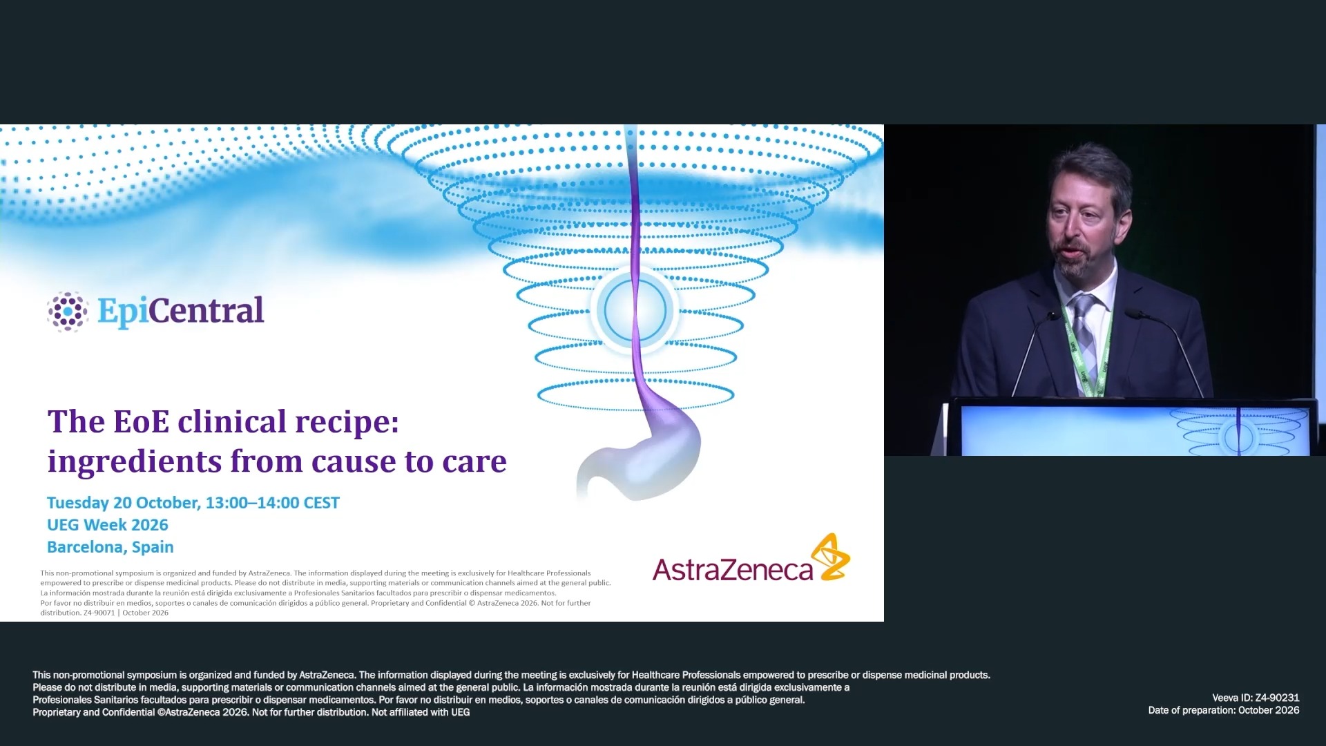
The EoE clinical recipe: ingredients from cause to care (AstraZeneca) (Complete Session)
Affiliations
1 University of North Carolina School of Medicine, Chapel Hill, NC, United States of America
2 USZ Zürich, Zurich, Switzerland
3 Otto-Von Guericke University, Madgeburg, Germany
4 Humanitas Gavazzeni e Castelli, None, Italy
5 Hospital General de Tomelloso, Madrid, Spain
Log in to continue.
This content is part of Gutflix. Log in with your myUEG account, or create one free, to watch it.
Not sure what you can access? Learn more about account types.
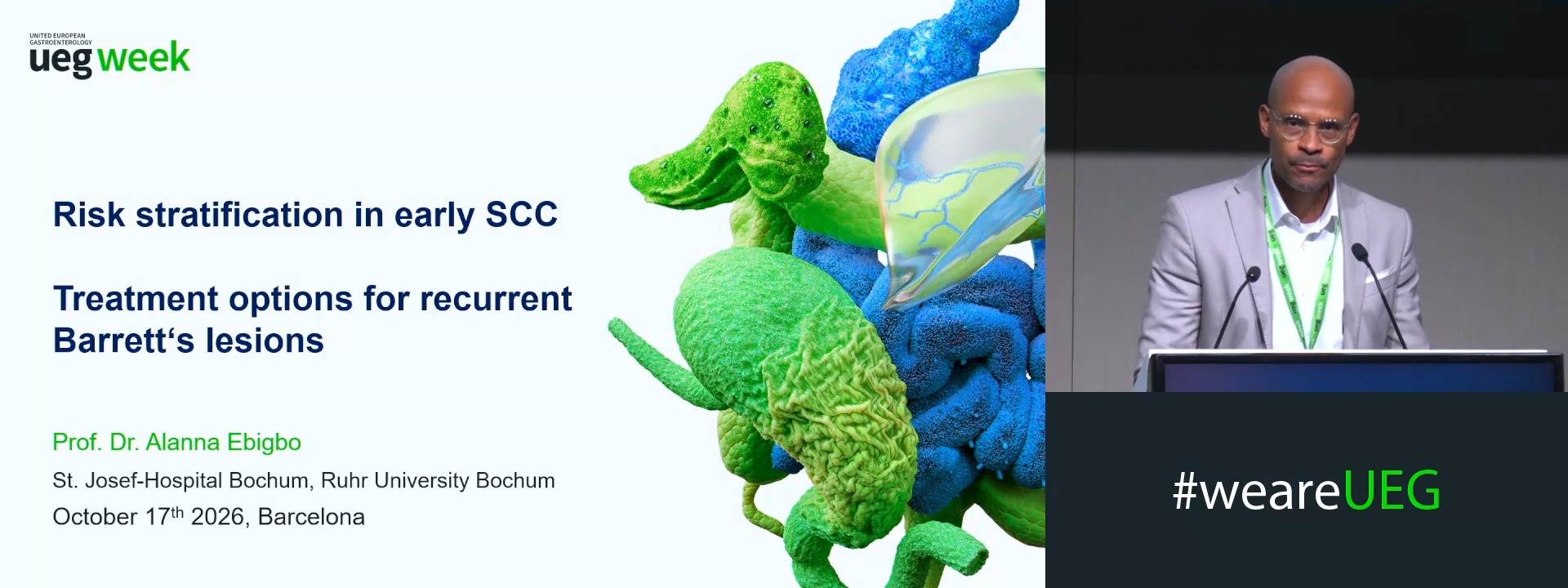
Risk stratification in early squamous cell carcinoma and treatment options for Barrett’s recurrence
Affiliations
1 St. Josef-Hospital, Ruhr-University Bochum, Bochum, Germany
Log in to continue.
This content is part of Gutflix. Log in with your myUEG account, or create one free, to watch it.
Not sure what you can access? Learn more about account types.
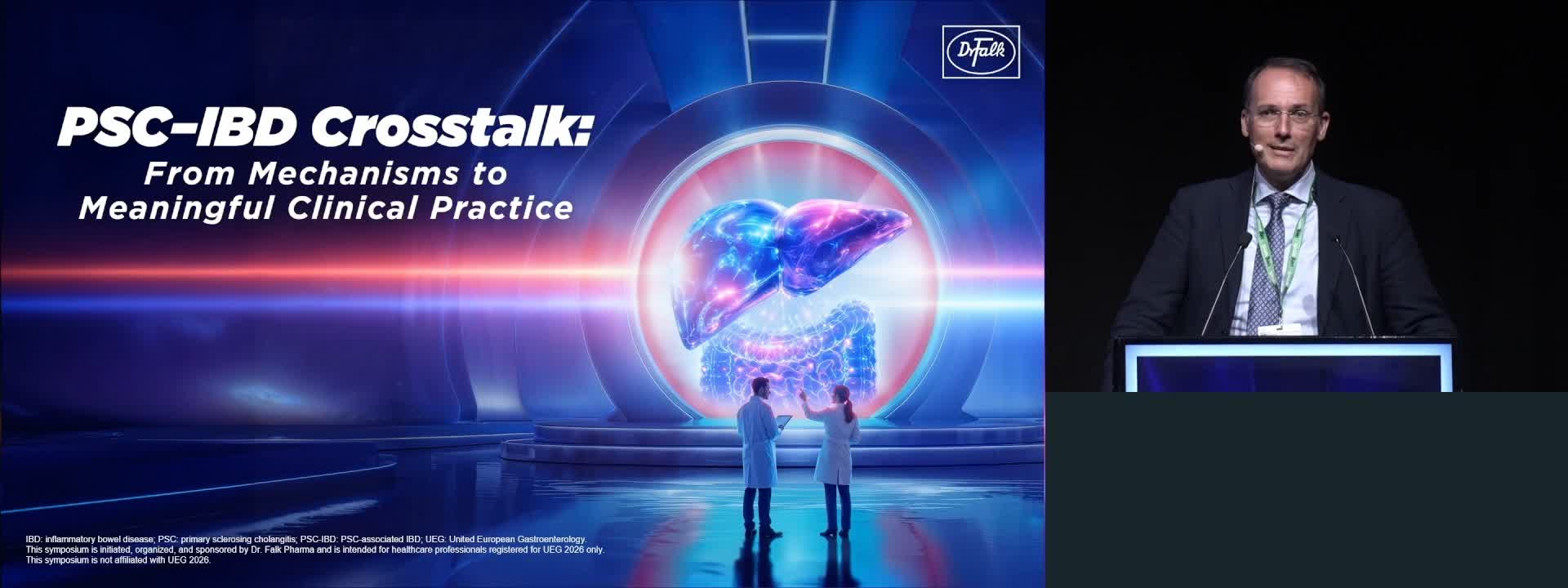
PSC-IBD crosstalk: From mechanisms to meaningful clinical practice (Dr. Falk Pharma) (Complete Session)
Affiliations
1 Charité - Universitätsmedizin, Berlin, Germany
2 Medical University of Vienna, Vienna, Austria
3 John Radcliffe Hospital, Oxford, Oxford, United Kingdom
4 University of Birmingham, Birmingham, United Kingdom

Log in to continue.
This content is part of Gutflix. Log in with your myUEG account, or create one free, to watch it.
Not sure what you can access? Learn more about account types.
How to evaluate abnormal liver biochemistry in asymptomatic people
Affiliations
1 New Castle Univerity, New Castle, United Kingdom