Introduction
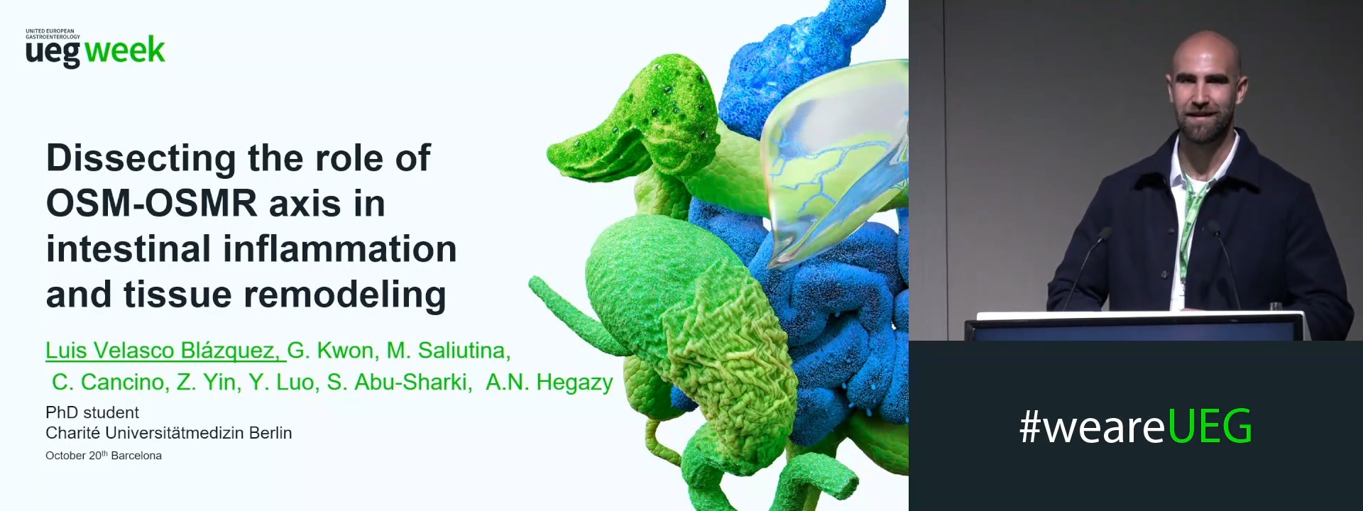
Oncostatin M (OSM) tracks with intestinal inflammation and predicts poor treatment response in inflammatory bowel disease (IBD). Chronic inflammation in patients presents with varied histological and molecular features, and repeated tissue injury drives fibrosis and aberrant remodeling. How the OSM-OSMR axis contributes across these different pathological contexts, and whether its role depends on the type of tissue injury and the cellular compartment that responds to OSM, remains poorly understood.
Aims & Methods
We examined the relationship between OSM-associated inflammatory molecular signatures and tissue pathology in human IBD, then tested the functional role of the OSM-OSMR axis in experimental intestinal inflammation. Human intestinal biopsies and surgical resections were stratified by an inflammatory gene module containing OSM into Inflammatory module high and low groups, then compared with healthy controls. We assessed tissue features with PathAI-based digital pathology and spatial transcriptomics, integrated with bulk RNAseq-derived inflammatory profiles. To mimic various type of tissue injury, we used DSS-induced colitis to model epithelial barrier disruption, testing global and cell-specific OSM-OSMR deficient mice across acute and chronic DSS inflammation and fibrosis with histopathological scoring, inflammatory gene signatures and flow cytometry of immune and stromal compartments.
Results
OSM Inflammatory module high patients displayed increased histopathological score, epithelial erosions and ulcerations compared with Inflammatory module low patients, together with increased lamina propria neutrophil infiltration. Spatial analysis highlighted spatial proximity of OSMhi neutrophils to epithelial erosions and ulcerations. Despite these associations, and the protective effect of OSMR depletion previously described, disrupting OSM-OSMR signaling across multiple cellular compartments did not alter histopathological severity in this model of acute epithelial loss. Compartment-dependent phenotypes emerged instead during chronic DSS. Endothelial OSMR deletion altered collagen deposition without affecting histopathological inflammation or colon features after three cycles of DSS. Conversely, OSMR deletion in stromal cells preserved colon length and reduced immune-cell infiltration, suggesting mesenchymal OSMR signaling contributes to chronic intestinal inflammation.
Conclusion
An OSM-associated inflammatory program identifies IBD with severe epithelial damage and increased neutrophil infiltration. OSM-OSMR disruption does not consistently improve acute epithelial injury. In chronic DSS, OSMR signaling in distinct compartments contributes to different features of disease, with endothelial OSMR linked to tissue remodeling and mesenchymal OSMR to immune infiltration. Together, these findings show that the functional consequences of OSM-OSMR signaling depend on pathological features, disease stage and the responding cellular compartment.
References
1. West, N. R., Hegazy, A. N., Owens, B. M., Bullers, S. J., Linggi, B., Buonocore, S., ... & Powrie, F. (2017). Oncostatin M drives intestinal inflammation and predicts response to tumor necrosis factor–neutralizing therapy in patients with inflammatory bowel disease. Nature medicine, 23(5), 579-589.
2. Cineus, R., Luo, Y., Saliutina, M., Manna, S., Cancino, C. A., Velasco Blázquez, L., ... & Hegazy, A. N. (2025). The IL-22–oncostatin M axis promotes intestinal inflammation and tumorigenesis. Nature immunology, 26(6), 837-853.
3. West, N. R., Owens, B. M., & Hegazy, A. N. (2018). The oncostatin M‐stromal cell axis in health and disease. Scandinavian journal of immunology, 88(3), e12694.