UEG's content platform for gastroenterology education bringing recordings, courses, guidelines, and more together in one place.
Trending on Gutflix
Rate for better recommendations!
Your feedback helps us show you more of what you like and less of what you dislike!
Log in to continue.
This content is part of Gutflix. Log in with your myUEG account, or create one free, to watch it.
Not sure what you can access? Learn more about account types.
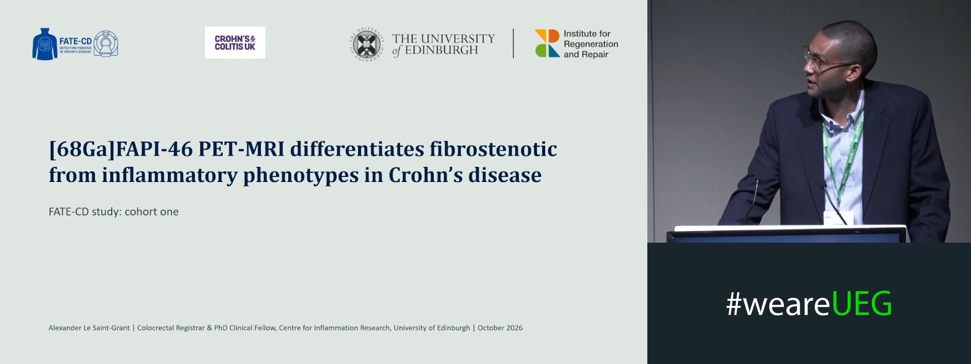
[68GA]FAPI PET-MRI QUANTIFIES FIBROBLAST ACTIVITY BURDEN AND DIFFERENTIATES FIBROSTENOSIS FROM AN INFLAMMATORY PHENOTYPE IN CROHN’S DISEASE
Alexander Le Saint-Grant 1,2, Solomon Xi Xiang Ong 1,2, Calum Worsley 3, Lucy Whitaker 1, Michelle Rooney 2, Nicholas T. Ventham 3, Scott Semple 2, Marc Dweck 2,4, Dilip Patel 3,2, Gwo-Tzer Ho 1,5, David Newby 2,4, Rahul Kalla 2,1
Affiliations
1 Institute for Regeneration and Repair, University of Edinburgh, Edinburgh, United Kingdom
2 Edinburgh Imaging Facility, University of Edinburgh, Edinburgh, United Kingdom
3 NHS Lothian, Edinburgh, United Kingdom
4 University of Edinburgh, Edinburgh, United Kingdom
5 University of Glasgow, Glasgow, United Kingdom
Log in to continue.
This content is part of Gutflix. Log in with your myUEG account, or create one free, to watch it.
Not sure what you can access? Learn more about account types.
Beyond borders: Managing a global nursing workforce – The impact of cultural diversity, beliefs, and training backgrounds on endoscopy services
Affiliations
1 Mater Misericordiae University Hospital, Dublin, Ireland
2 None, None, Switzerland
3 Mater Dei Hospital, Zabbar, Malta
Log in to continue.
This content is part of Gutflix. Log in with your myUEG account, or create one free, to watch it.
Not sure what you can access? Learn more about account types.
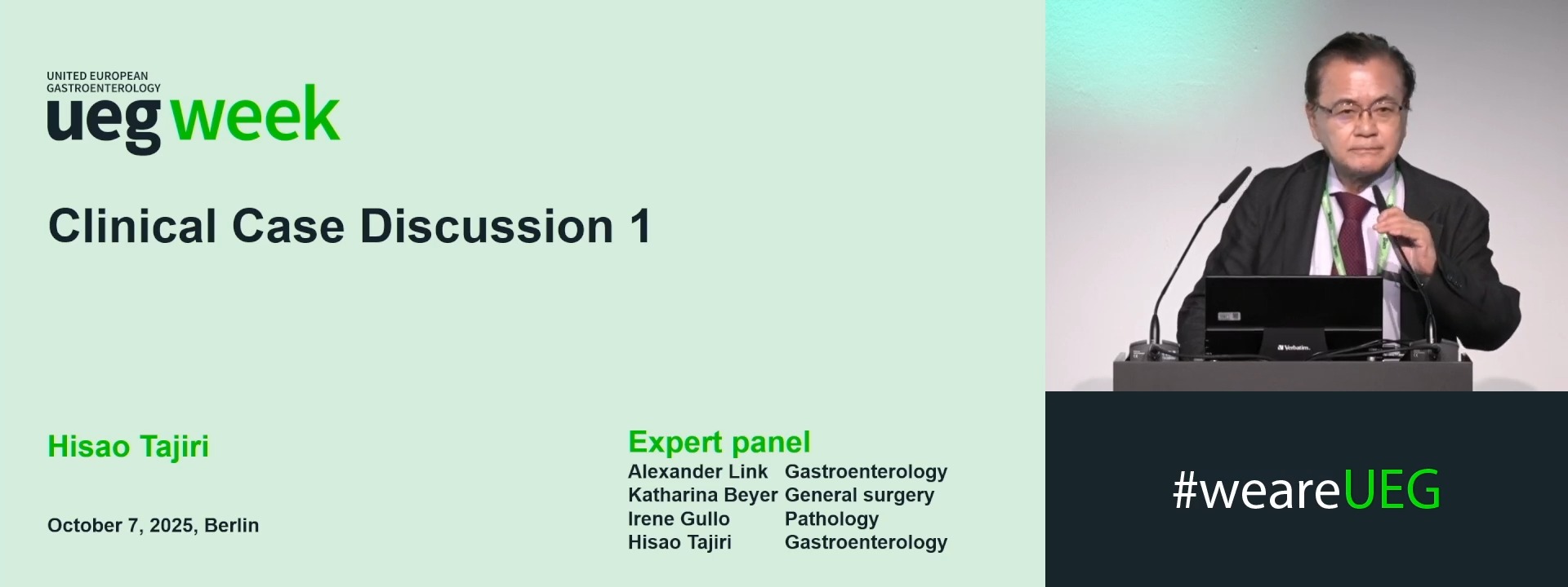
Early gastric cancer (Complete Session)