UEG's content platform for gastroenterology education bringing recordings, courses, guidelines, and more together in one place.
Trending on Gutflix
Rate for better recommendations!
Your feedback helps us show you more of what you like and less of what you dislike!
Log in to continue.
This content is part of Gutflix. Log in with your myUEG account, or create one free, to watch it.
Not sure what you can access? Learn more about account types.
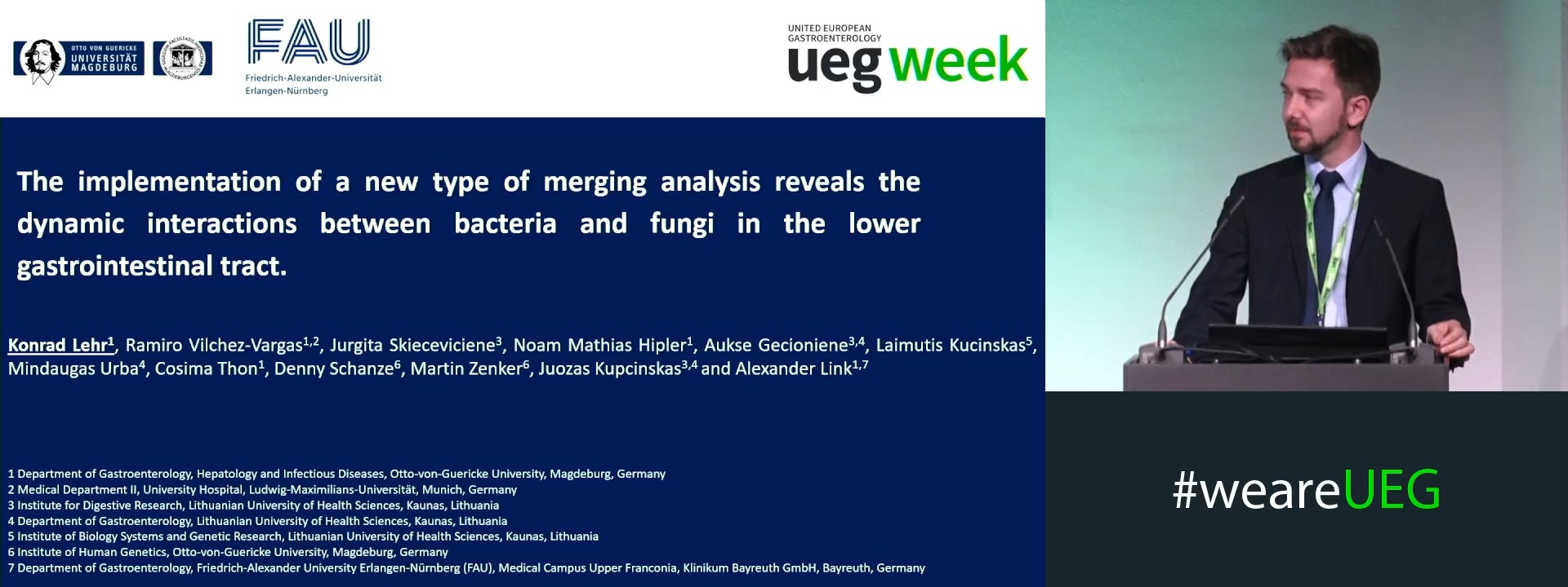
THE IMPLEMENTATION OF A NEW TYPE OF MERGING ANALYSIS REVEALS THE DYNAMIC INTERACTIONS BETWEEN BACTERIA AND FUNGI IN THE LOWER GASTROINTESTINAL TRACT
Konrad Lehr 1, Ramiro Vilchez-Vargas 1,2, Jurgita Skieceviciene 3, Noam Hipler 1, Aukse Gecioniene 3, Laimutis Kucinskas 3, Mindaugas Urba 3, Cosima Thon 1, Denny Schanze 1, Unknown Unknown 1, Juozas Kupcinskas 3, Alexander Link 1
Affiliations
1 Otto-von-Guericke University, Magdeburg, Germany
2 Ludwig-Maximilians-University, Munich, Germany
3 Lithuanian University of Health Sciences, Kaunas, Lithuania

Log in to continue.
This content is part of Gutflix. Log in with your myUEG account, or create one free, to watch it.
Not sure what you can access? Learn more about account types.
THE FUNGAL COMMUNITY IN THE LOWER GASTROINTESTINAL TRACT OF MONOZYGOTIC AND DIZYGOTIC TWINS
Konrad Lehr 1, Ramiro Vilchez-Vargas 1, Jurgita Skieceviciene 2, Noam Hipler 1, Cosima Thon 1, Aukse Gecioniene 2, Denny Schanze 1, Unknown Unknown 1, Juozas Kupcinskas 2, Alexander Link 1
Affiliations
1 Otto-von-Guericke University, Magdeburg, Germany
2 University of Health Sciences, Kaunas, Lithuania

Log in to continue.
This content is part of Gutflix. Log in with your myUEG account, or create one free, to watch it.
Not sure what you can access? Learn more about account types.
Introduction: Mistakes in... Lower GI
Affiliations
1 Stadtspital Triemli, Zürich, Switzerland

Log in to continue.
This content is part of Gutflix. Log in with your myUEG account, or create one free, to watch it.
Not sure what you can access? Learn more about account types.
Introduction Mistakes in…: Lower GI
Affiliations
1 Stadtspital Triemli, Zürich, Switzerland

Log in to continue.
This content is part of Gutflix. Log in with your myUEG account, or create one free, to watch it.
Not sure what you can access? Learn more about account types.
Introduction: Mistakes in...Lower GI
Affiliations
1 Stadtspital Triemli, Zürich, Switzerland