Introduction
Inflammatory Bowel Disease (IBD)1,2 requires close monitoring to detect flare ups early and guide treatment. While endoscopy is the gold standard, it is invasive, costly, and uncomfortable1,2. Intestinal Ultrasound (IUS) is a non-invasive alternative with strong correlation to endoscopic findings—especially bowel wall thickness (BWT), a marker of disease activity3. Nevertheless, IUS remains limited due to operator dependence and a steep learning curve4.
AI has improved consistency in other domains, e.g. endoscopic evaluation, but its application to IUS in IBD remains underexplored. Prior models lack interpretability and require manual image cropping, or are trained on ideal, selected data, limiting clinical use5,6.
Aims & Methods
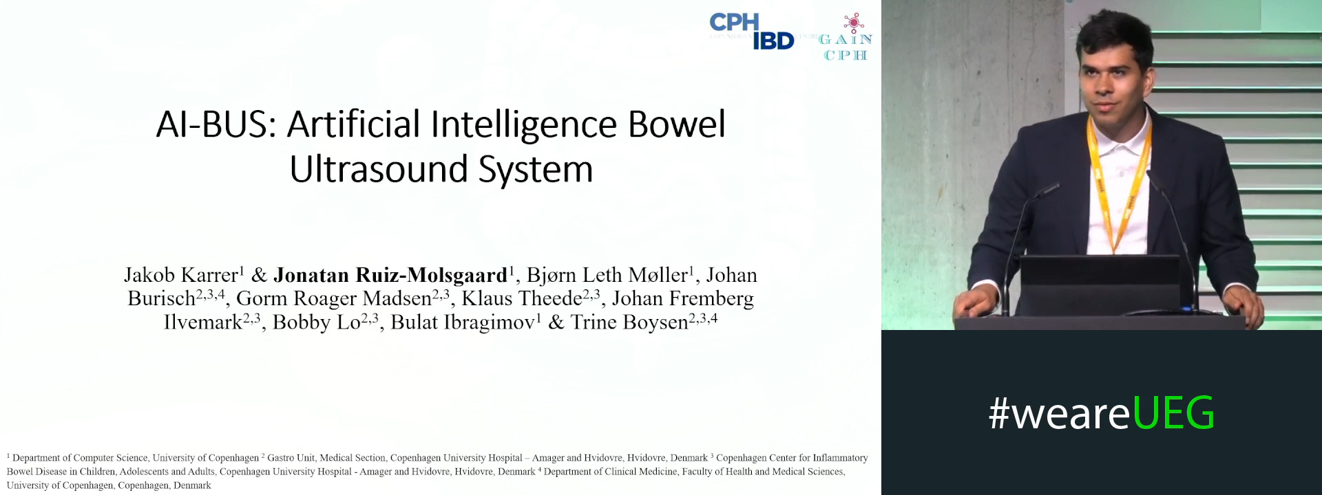
We aim to develop a deep-learning model that automatically identifies and paints the bowel wall and measures BWT directly from raw clinical IUS images.
A training dataset of 570 images from 144 IUS videos and a testing dataset of 55 images from 55 separate exams were created. All images were extracted from previously performed IUS examinations reflecting real-world variation, including e.g. unclear boundaries and artefacts.
All images were annotated by International Bowel Ultrasound Group (IBUS) certified experts, including outline-paintings of the inner and outer bowel wall and two BWT measurements.
The AI consists of a combination of Convolutional neural networks and other image processing algorithms.
Evaluation included BWT error against the expert mean, classification accuracy using the standard IBUS 3 mm threshold3, and a leave-one-out comparison with individual doctors.
Results
The model produced predictions on 54 of 55 test images.
The model deviated from the gold standard mean by 0.98 mm (SD 1.1 mm) per image on the regression task. The average distance to expert-defined bounds was 0.44 mm (SD 0.89 mm), with 59% of predictions inside this range.
For classification (using a 3 mm threshold), the model reached an accuracy of 0.77, sensitivity of 0.69, specificity of 0.94, and Cohen’s Kappa of 0.56.
In the leave-one-out analysis, expert performance ranged from 0.89–0.93 accuracy and 0.79–0.83 Kappa. Depending on which expert was excluded from the gold standard, the model achieved 0.74–0.80 accuracy and 0.49–0.60 Kappa. Experts stayed within expert-defined bounds in 72–81% of cases, while the model did so in 46–55%.
Conclusion
Clinically, the model performed well. An error of 0.5–1 mm is negligible in practice and matches typical variation in manual measurements. Many real-world test images had BWT values near the 3 mm threshold, so small deviations led to misclassifications. In practice, a 2.5 mm reading could still raise concerns based on symptoms3.
While the system alone cannot provide expert-level BWT measurements, it could provide assistance to experts as well as non-expert and junior doctors, especially in locating the bowel, which is an essential part of IUS.
The model works on unprocessed clinical data, with the only selection criteria being that there is an identifiable bowel segment, measurable by an IBUS-certified doctor. Allowing the images to reflect realistic conditions
In summary, we developed an AI model that identifies the bowel and measures BWT with acceptable accuracy. We have already started the processes of collecting video data, to extend the AI’s functionality to a fully clinical setting.
References
1. Torres J, Mehandru S, Colombel JF, Peyrin-Biroulet L. Crohn’s disease. The Lancet. 2017;389(10080):1741-1755. doi:10.1016/S0140-6736(16)31711-1
2. Ungaro R, Mehandru S, Allen PB, Peyrin-Biroulet L, Colombel JF. Ulcerative colitis. Lancet. 2017;389(10080):1756-1770. doi:10.1016/S0140-6736(16)32126-2
3. Novak KL, Group I, Nylund K, et al. Expert Consensus on Optimal Acquisition and Development of the International Bowel Ultrasound Segmental Activity Score [IBUS-SAS]: A Reliability and Inter-rater Variability Study on Intestinal Ultrasonography in Crohn’s Disease. J Crohns Colitis. 2021;15(4):609-616. doi:10.1093/ECCO-JCC/JJAA216
4 Bove L, Meyer J, Collins M, Frampas E, Bourreille A, Le Berre C. Understanding the learning curve of intestinal ultrasound in inflammatory bowel disease: A comparative study between novice, regular, and expert. Clin Res Hepatol Gastroenterol. 2025;49(3):102548. doi:10.1016/J.CLINRE.2025.102548
5. Kumaralingam L, Le May K, Dang VB, Alavi J, Huynh HQ, Le LH. Artificial intelligence-assisted approach to assessing bowel wall thickness in pediatric inflammatory bowel disease using intestinal ultrasound images. J Crohns Colitis. 2025;19(4). doi:10.1093/ECCO-JCC/JJAF037
6. Carter D, Albshesh A, Shimon C, et al. Automatized Detection of Crohn’s Disease in Intestinal Ultrasound Using Convolutional Neural Network. Inflamm Bowel Dis. February 2023:1-6. doi:10.1093/IBD/IZAD014