UEG's content platform for gastroenterology education bringing recordings, courses, guidelines, and more together in one place.
Trending on Gutflix
Rate for better recommendations!
Your feedback helps us show you more of what you like and less of what you dislike!
Log in to continue.
This content is part of Gutflix. Log in with your myUEG account, or create one free, to watch it.
Not sure what you can access? Learn more about account types.
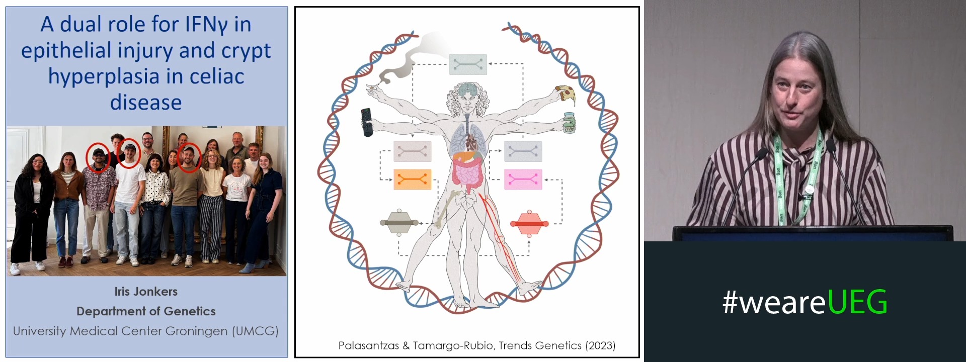
A DUAL ROLE FOR IFNG IN EPITHELIAL INJURY AND CRYPT HYPERPLASIA IN CELIAC DISEASE
Affiliations
1 UMCG, Groningen, Netherlands
Log in to continue.
This content is part of Gutflix. Log in with your myUEG account, or create one free, to watch it.
Not sure what you can access? Learn more about account types.
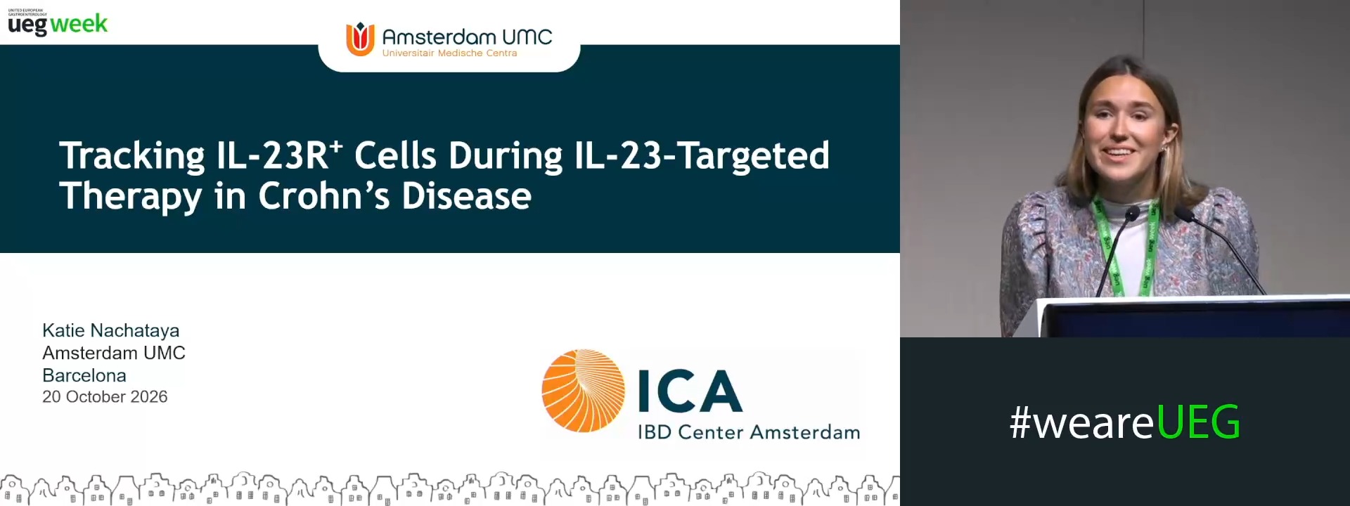
SINGLE-CELL RNA SEQUENCING REVEALS TRANSCRIPTIONAL REMODELLING OF IL-23R⁺ CELLS FOLLOWING IL-23–TARGETED THERAPY IN PATIENTS WITH CROHN’S DISEASE AFTER ILEOCAECAL RESECTION
Affiliations
1 Amsterdam UMC, Amsterdam, Netherlands
2 VUMC, Amsterdam, Netherlands
3 AMC Amsterdam Inflammatory Bowel Disease Centre - Academic Medical Center, AMC Amsterdam Inflammator, Amsterdam, Netherlands
4 Academisch Medisch Centrum, Amsterdam, Netherlands
Log in to continue.
This content is part of Gutflix. Log in with your myUEG account, or create one free, to watch it.
Not sure what you can access? Learn more about account types.
Summary: Dilemmas in ulcerative colitis: Back to the basics or fast forward
Affiliations
1 John Radcliffe Hospital, Oxford, United Kingdom
Log in to continue.
This content is part of Gutflix. Log in with your myUEG account, or create one free, to watch it.
Not sure what you can access? Learn more about account types.
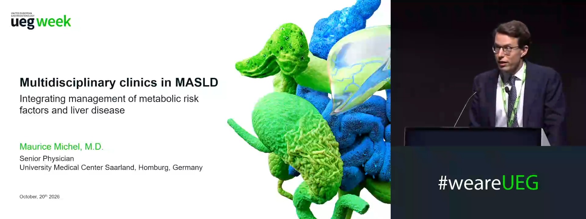
Multidisciplinary clinics in MASLD: Integrating management of metabolic risk factors and liver disease
Affiliations
1 Department of Internal Medicine II, University Medical Center Saarland, Homburg, Germany