UEG's educational platform for gastroenterology education bringing recordings, courses, guidelines, and more together in one place.
Most popular content
Rate for better recommendations!
Your feedback helps us show you more of what you like and less of what you dislike!
Log in to access this content.
Free for all myUEG account holders. Your access level is set automatically based on your occupation. Medical professionals get full access to all content. If you are a non-medical user, you can only access UEG Week content from congresses you attended.
Not sure what you can access? Learn more about account types.

IS THERE AN ASSOCIATION BETWEEN CELIAC DISEASE AND HELICOBACTER PYLORI?
AZOUZOUT CHAMS 1, ikram douhri 2, Ouiam Elmqaddem 2, Hajar Koulali 2, Zazour Abdelkrim 2, Zahi Ismaili 2, Ghizlane Kharrasse 2
1 Faculty of Medicine and Pharmacy, Mohammed First University, Oujda, Morocco|||Mohammed VI University Hospital Center, Oujda, Morocco
2 Faculty of Medicine and Pharmacy, Mohammed First University, Oujda, Morocco
Conference
Topics
Submission format
Session
Citation
Published
Log in to access this content.
Free for all myUEG account holders. Your access level is set automatically based on your occupation. Medical professionals get full access to all content. If you are a non-medical user, you can only access UEG Week content from congresses you attended.
Not sure what you can access? Learn more about account types.
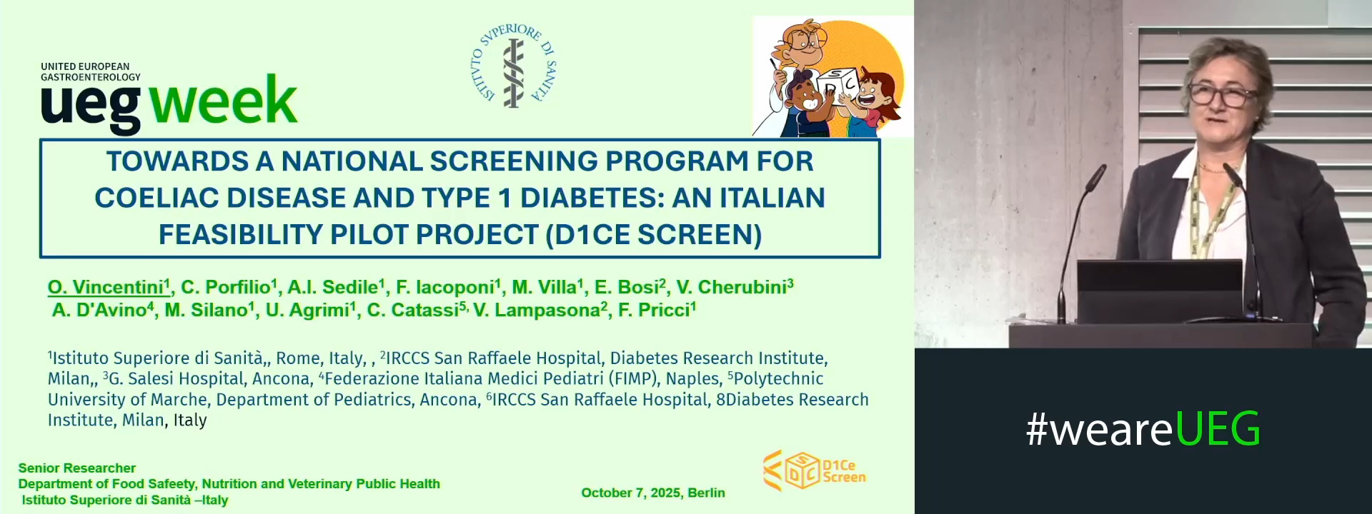
TOWARDS A NATIONAL SCREENING PROGRAM FOR COELIAC DISEASE AND TYPE 1 DIABETES: AN ITALIAN FEASIBILITY PILOT PROJECT (D1CE SCREEN)
Olimpia Vincentini 1, Chiara Porfilio 1, Adalgisa Ilaria Sedile 1, Francesca Iacoponi 1, Marika Villa 1, Emanuele Bosi 2, Valentino Cherubini 3, Antonio D'Avino 4, Marco Silano 1, Umberto Agrimi 1, Carlo Catassi 5, Vito Lampasona 2, Flavia Pricci 1
1 Istituto Superiore di Sanità, Rome, Italy
2 IRCCS San Raffaele Hospital, Milan, Italy
3 Azienda Ospedaliero-Universitaria, Ospedali Riuniti di Ancona, G. Salesi Hospital, Ancona, Italy
4 Federazione Italiana Medici Pediatri (FIMP), Naples, Italy
5 Polytechnic University of Marche, Ancona, Italy
Event
Submission format
Citation
Published
Log in to access this content.
Free for all myUEG account holders. Your access level is set automatically based on your occupation. Medical professionals get full access to all content. If you are a non-medical user, you can only access UEG Week content from congresses you attended.
Not sure what you can access? Learn more about account types.
Introduction
Interpretation of duodenal biopsies in celiac disease (CeD) is receiving attention in clinical practice and clinical trials, where follow up biopsies are obtained to assess CeD activity or response to interventions. Follow up biopsies are likely to increase once therapeutics are approved, to determine eligibililty. While CeD research pathologists employ defined histologic protocols in grading celiac disease injury, diagnostic practices of non-CeD focused gastrointestinal pathologists(GI) are unknown.
Aims & Methods
A 19-question survey was distributed to 489 United States based GI pathologists. Topics included demographics, approach to diagnosis, reporting preferences and availability/attitudes towards digital pathology. A follow up survey of micrographs of duodenal mucosal biopsies was then distributed. Respondents were asked to comment on adequacy of mucosal orientation to assess architecture and to score villous blunting in celiac disease/control biopsies.
Results
There were143 responses (29%). See Table 1 for detailed responses for Demographics, Pathology Assessment, Reporting and Digital Pathology Status. 65% evaluate >25 biopsies per week. Quality of biopsy orientation was defined by 87% of respondents as the presence of at least 3 oriented villous-crypt units. 76% stated that inadequate orientation occurs in only a minority of samples (<25%), yet the ability of respondents to recognize poorly oriented biopsies was limited in the examples shown in the follow up survey. When presented with histology images, 93% of respondents correctly stated that a well-oriented example biopsy was adequate to evaluate villous architecture. However, only 24% of respondents recognized that a second image with no evaluable villous-crypt units was inadequately oriented, and would therefore be unevaluable for grading villous architecture. 15% review existing prior biopsy slides routinely, while 46% review existing prior biopsies when specifically requested to do so. Respondent inter-observer agreement on villous blunting scores on biopsy images was best in the setting of normal mucosa or severe villous blunting, and decreased in the setting of mild to moderate blunting. 44% would welcome software assisted digital sign out with villous height: crypt depth and intraepithelial lymphocytes (IEL) measuring tools, provided it would not increase sign out time by more than 1-2 minutes per case.
Table 1:
| Question Category | Question: 143/489 (29% response rate) | Results: Number (%) |
| Demographics of Respondents | Gastrointestinal Pathology Fellowship Trained | 106 (74) |
| Academic Practice Environment | 101 (71) | |
| Biopsy Orientation | Define adequate biopsy orientation as at least 3 oriented villous-crypt units | 116 (87) |
| I always request level sections if biopsies are inadequately oriented | 20 (15) | |
| Diagnostic Approach | Provide Marsh-Oberhuber score in reports | 32 (24) |
| Describe range of villous blunting/IELs, including whether diffuse or patchy | 101 (77) | |
| Evaluate IELs on H&E stains only | 114 (86) | |
| Digital Status of Practice | Currently employ digital sign out in practice | 3 (2) |
| Anticipate digital sign out within 10 years | 52 (39) |
Conclusion
Among 143 US GI pathologists, there is significant heterogeneity in CeD pathology reporting. Respondents likely overestimate the adequacy of biopsy orientation in routine sections, which affects assessments of villous architecture. Interobserver variation was noted in architecture scores in 6 examples. Digital pathology has not been implemented in most practices. Challenges and resistance to employing online measuring tools was expressed. These data reveal opportunities for improvement in pathologist's approach to CeD biopsy interpretation.
References
Robert ME, Ciacci C, Lebwhol B. Gastroenterology. 2024 PMID: 38302007.
Disclosure
Dr. Robert consults for Takeda, Anokion, Teva, Alimentiv, PathAI and receives research salary support from Astra-Zeneca. Dr. Murray consults for Cour, Takeda and Johnson and Johnson. Dr. Leffler receives salary support from Takeda.
CURRENT PRACTICES AND OPPORTUNITIES IN HISTOLOGIC INTERPRETATION OF CELIAC DISEASE DUODENAL BIOPSIES IN THE PERI-DIGITAL ERA
1 Yale University School Of Medicine, New Haven, United States
2 Mayo Clinic, Rochester, United States
3 Beth Israel Deaconness Medical Center, Boston, United States
Conference
Topics
Submission format
Citation
Published
Log in to access this content.
Free for all myUEG account holders. Your access level is set automatically based on your occupation. Medical professionals get full access to all content. If you are a non-medical user, you can only access UEG Week content from congresses you attended.
Not sure what you can access? Learn more about account types.
FROM POTENTIAL TO OVERT CELIAC DISEASE: IDENTIFYING WHO IS AT RISK AND WHEN PROGRESSION OCCURS
Elia Fracas 1, Elena Del Zanna 1, Nicoletta Nandi 2, Lorenzo Norsa 3, Agostino Cosenza 1, Lucia Scaramella 4, Luisa Lonoce 5, Matilde Topa 1, Leda Roncoroni 1, Alice Scricciolo 4, Vincenza Lombardo 4, Cristina Cocuccio 3, Elena Pozzi 3, Beatrice Rota 4, Luca Elli 1
1 University of Milano, Milano, Italy
2 Sheffield Teaching Hospital, Sheffield, United Kingdom
3 Ospedale dei Bambini “Vittore Buzzi”, Milano, Italy
4 Fondazione IRCCS Ca' Granda Ospedale Maggiore Policlinico, Milan, Italy
5 Azienda Ospedaliera Universitaria Senese, Siena, Italy
Conference
Topics
Submission format
Citation
Published
Log in to access this content.
Free for all myUEG account holders. Your access level is set automatically based on your occupation. Medical professionals get full access to all content. If you are a non-medical user, you can only access UEG Week content from congresses you attended.
Not sure what you can access? Learn more about account types.
IS IT USEFUL TO MEASURE QUALITY OF LIFE IN THE FOLLOW-UP OF PATIENTS WITH CELIAC DISEASE? A SINGLE-CENTER EXPERIENCE
1 Hospital Universitario Virgen de las Nieves, Granada, Spain
Conference
Topics
Submission format
Session
Citation
Published
Log in to access this content.
Free for all myUEG account holders. Your access level is set automatically based on your occupation. Medical professionals get full access to all content. If you are a non-medical user, you can only access UEG Week content from congresses you attended.
Not sure what you can access? Learn more about account types.
ANALYTICAL COMPARISON OF A NOVEL, FULLY AUTOMATED MULTIPLEXED MICROARRAY IMMUNOASSAY AND ROUTINE METHODS FOR THE DETECTION OF CELIAC DISEASE ANTIBODIES IN A LABORATORY IN ENGLAND
1 Peterborough City Hospital, North West Anglia NHS Foundation Trust, Peterborough, United Kingdom
2 AliveDx, Eysins, Switzerland
Conference
Topics
Submission format
Session
Citation
Published
Log in to access this content.
Free for all myUEG account holders. Your access level is set automatically based on your occupation. Medical professionals get full access to all content. If you are a non-medical user, you can only access UEG Week content from congresses you attended.
Not sure what you can access? Learn more about account types.
COELIAC DISEASE IN BONE FRACTURE PATIENTS: A 1000 PATIENT PILOT SCREENING STUDY
Frankie Fan 1, Alex Calder 2, Sophie Fox 2, Parizade Raymode 2, Richard Harris 2, Srinivasan Shyamsundar 2, Ajay Verma 2
1 Kettering General Hospital, Kettering, United Kingdom|||University of Leicester, Leicester, United Kingdom
2 Kettering General Hospital, Kettering, United Kingdom
Conference
Topics
Submission format
Session