UEG's content platform for gastroenterology education bringing recordings, courses, guidelines, and more together in one place.
Trending on Gutflix
Rate for better recommendations!
Your feedback helps us show you more of what you like and less of what you dislike!
Log in to continue.
This content is part of Gutflix. Log in with your myUEG account, or create one free, to watch it.
Not sure what you can access? Learn more about account types.
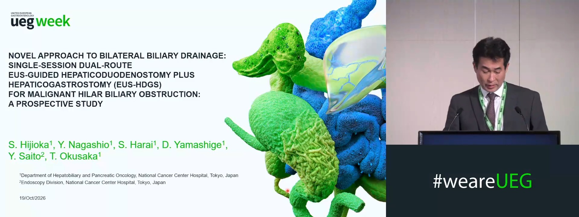
NOVEL APPROACH TO BILATERAL BILIARY DRAINAGE: SINGLE-SESSION DUAL-ROUTE EUS-GUIDED HEPATICODUODENOSTOMY PLUS HEPATICOGASTROSTOMY FOR MALIGNANT HILAR BILIARY OBSTRUCTION: A PROSPECTIVE STUDY
SUSUMU HIJIOKA 1, Yoshikuni Nagashio 1, Shota Harai 1, Daiki Yamashige 1, Yutaka Saito 2, Takuji Okusaka 1
Affiliations
1 National Cancer Center Hospital, Tokyo, Japan
2 National Cancer Center Hospital Endoscopy Division, Tokyo, Japan
Log in to continue.
This content is part of Gutflix. Log in with your myUEG account, or create one free, to watch it.
Not sure what you can access? Learn more about account types.
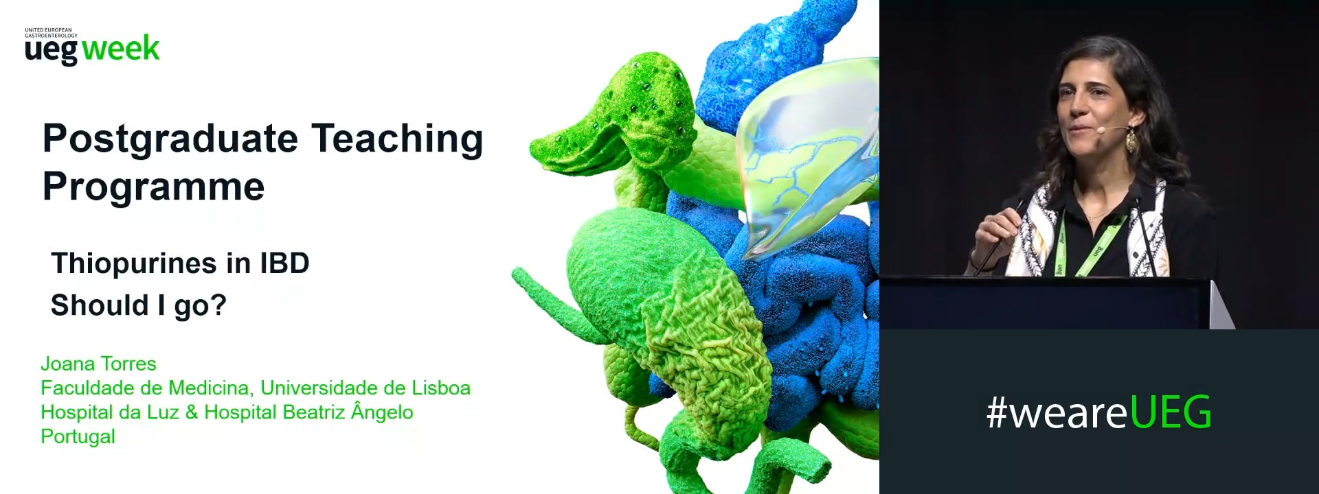
Current role for thiopurines in the management of IBD: Should I go?
Affiliations
1 Hospital Beatriz Ângelo & Hospital da Luz, Lisbon, Portugal
Log in to continue.
This content is part of Gutflix. Log in with your myUEG account, or create one free, to watch it.
Not sure what you can access? Learn more about account types.
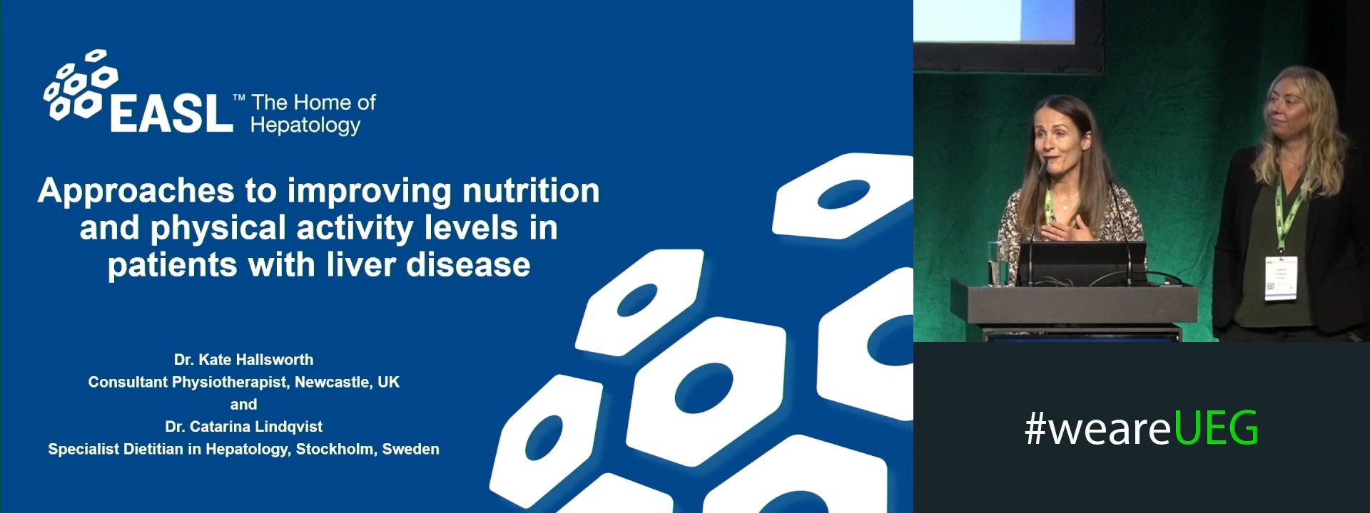
Approaches to improve nutrition and physical activity levels in patients with liver disease
Affiliations
1 NIHR Newcastle Biomedical Research Centre, Newcastle upon Tyne, United Kingdom
2 None, None, Sweden
Log in to continue.
This content is part of Gutflix. Log in with your myUEG account, or create one free, to watch it.
Not sure what you can access? Learn more about account types.
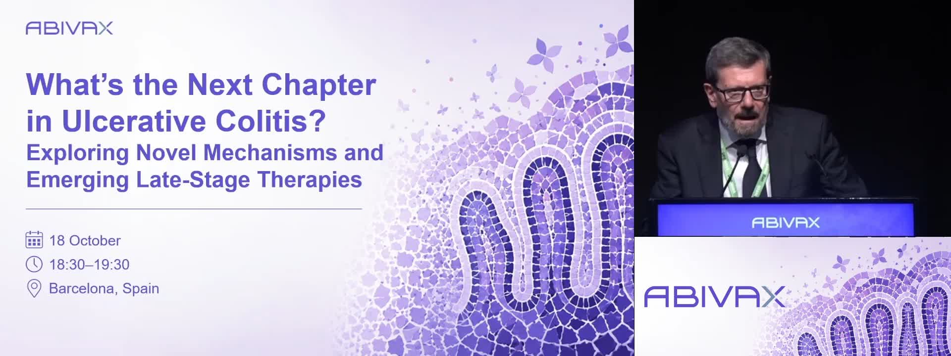
What’s the Next Chapter in Ulcerative Colitis? Exploring Novel Mechanisms and Emerging Late-Stage Therapies (Abivax) (Complete Session)
Affiliations
1 University of Calgary, Calgary, Canada
2 IBD Center, IRCCS Humanitas Research Hospital, Rozzano, Milan, Italy
3 Friedrich-Alexander-Universität Erlangen-Nürnberg, Erlangen, Germany
Log in to continue.
This content is part of Gutflix. Log in with your myUEG account, or create one free, to watch it.
Not sure what you can access? Learn more about account types.
ASSESSING THE LIMITS OF ENDOSCOPIC ULTRASONOGRAPHY IN DETECTING PANCREATIC MICROARCHITECTURE: A SIDE-BY-SIDE COMPARISON WITH SURGICAL SPECIMENS
Jun Nakahodo 1,2, Kensuke Hoshi 1,3, Shin-ichiro Horiguchi 1, Yasuji Seyama 1, Hiroko Okinaga 1, Mei Nakamura 1, Tomoaki Sawano 1, Masanao Kurata 1, Kazuro Chiba 1, Hiroki Tabata 1, WATARU UJITA 1, Ryogo Minami 1, Sorano Ichiya 1, T. Kamisawa 1, TOSHIRO IIZUKA 1
Affiliations
1 Tokyo Metropolitan Cancer and Infectious Diseases Center, Komagome Hospital, Bunkyo-Ku, Japan
2 Juntendo University, Bunkyo-Ku, Japan
3 Toho University Omori Medical Center, Ota-ku, Japan