UEG's educational platform for gastroenterology education bringing recordings, courses, guidelines, and more together in one place.
Most popular content
Rate for better recommendations!
Your feedback helps us show you more of what you like and less of what you dislike!
Log in to access this content.
Free for all myUEG account holders. Your access level is set automatically based on your occupation. Medical professionals get full access to all content. If you are a non-medical user, you can only access UEG Week content from congresses you attended.
Not sure what you can access? Learn more about account types.
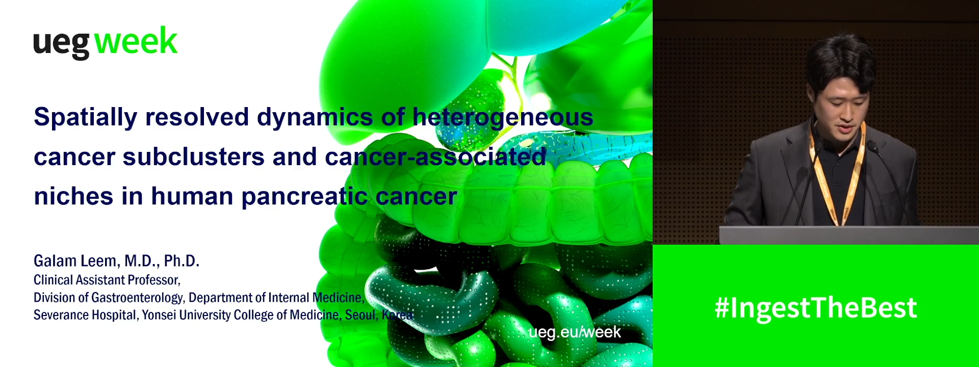
SPATIALLY RESOLVED DYNAMICS OF HETEROGENEOUS CANCER SUBCLUSTERS AND CANCER-ASSOCIATED NICHES IN HUMAN PANCREATIC CANCER
1 Yonsei University College of Medicine, Seoul, Korea, Republic of
Event
Submission format
Citation
Published
Log in to access this content.
Free for all myUEG account holders. Your access level is set automatically based on your occupation. Medical professionals get full access to all content. If you are a non-medical user, you can only access UEG Week content from congresses you attended.
Not sure what you can access? Learn more about account types.
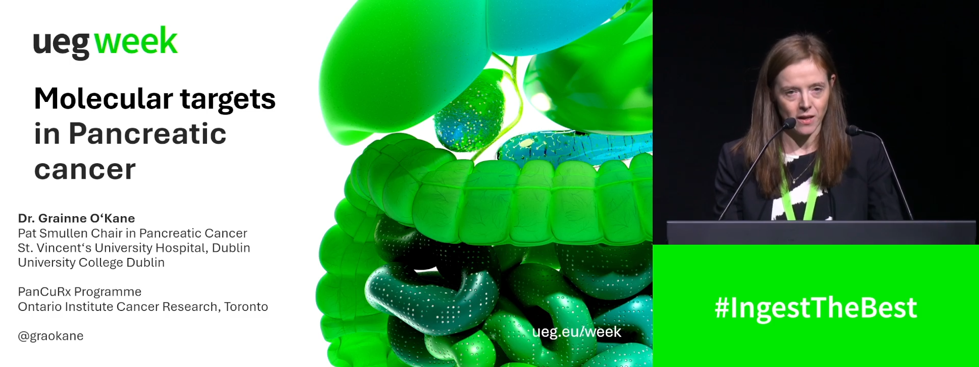
Molecular targets in pancreatic cancer
1 Trinity St James's Cancer Institute, Dublin, Ireland
Event
Topics
Citation
Published
Log in to access this content.
Free for all myUEG account holders. Your access level is set automatically based on your occupation. Medical professionals get full access to all content. If you are a non-medical user, you can only access UEG Week content from congresses you attended.
Not sure what you can access? Learn more about account types.
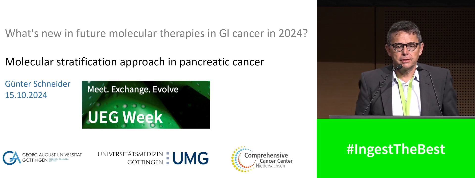
Molecular stratification approach in pancreatic cancer
1 Universitätsmedizin Göttingen, Göttingen, Germany
Event
Citation
Published
Log in to access this content.
Free for all myUEG account holders. Your access level is set automatically based on your occupation. Medical professionals get full access to all content. If you are a non-medical user, you can only access UEG Week content from congresses you attended.
Not sure what you can access? Learn more about account types.
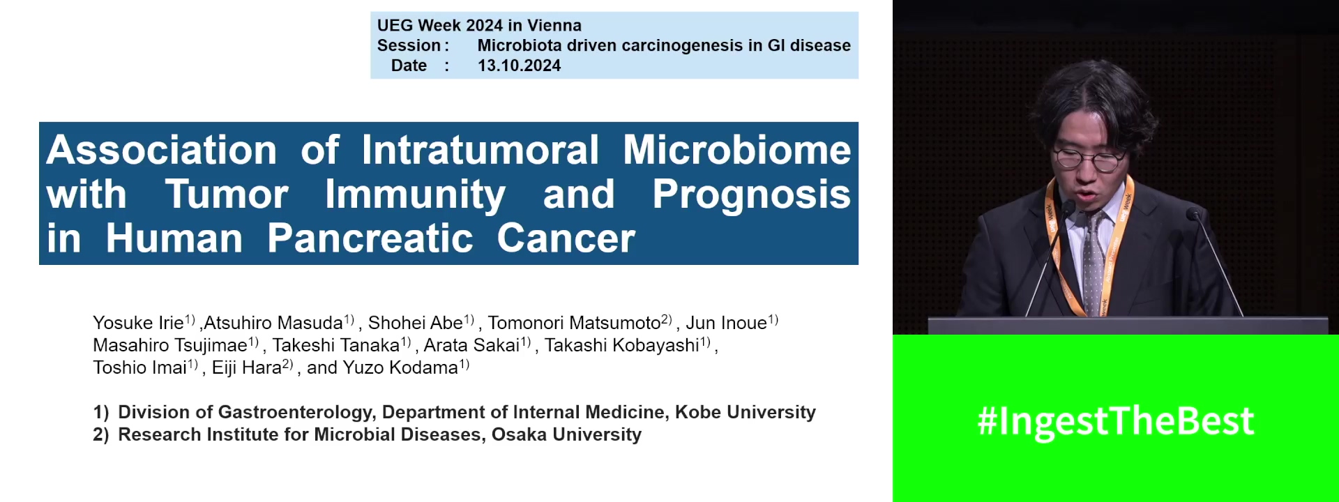
ASSOCIATION OF INTRATUMORAL MICROBIOME WITH TUMOR IMMUNITY AND PROGNOSIS IN HUMAN PANCREATIC CANCER
Yosuke Irie 1, Atsuhiro Masuda 1, Shohei Abe 1, Tomonori Matsumoto 2, Jun Inoue 1, Masahiro Tsujimae 1, Takeshi Tanaka 1, Arata Sakai 1, Takashi Kobayashi 1, Toshio Imai 1, Eiji Hara 3, Yuzo Kodama 1
1 Kobe graduate school of medicine, Kobe, Japan
2 Osaka University, Suita, Japan
3 Osaka University, Osaka, Japan
Event
Submission format
Citation
Published
Log in to access this content.
Free for all myUEG account holders. Your access level is set automatically based on your occupation. Medical professionals get full access to all content. If you are a non-medical user, you can only access UEG Week content from congresses you attended.
Not sure what you can access? Learn more about account types.
Molecular subgroups in pancreatic cancer for treatment selection
1 Klinikum Rechts Der Isar, Technical University of Munich, Muenchen, Germany
Event
Citation
Published
Log in to access this content.
Free for all myUEG account holders. Your access level is set automatically based on your occupation. Medical professionals get full access to all content. If you are a non-medical user, you can only access UEG Week content from congresses you attended.
Not sure what you can access? Learn more about account types.

FOLFIRINOX VERSUS GEMCITABINE/NAB-PACLITAXEL IN METASTATIC PANCREATIC CANCER: A REAL-WORLD DATA ANALYSIS INCLUDING NGS PANEL INSIGHTS
Jeehoon Kim 1, Galam Leem 1, Hee Seung Lee 1, Moon-Jae Chung 1, Jeong Youp Park 1, Seungmin Bang 1, Seung Woo Park 1, Si-Young Song 1, Jung Hyun Jo 1
1 Yonsei University College of Medicine, Seoul, Korea, Republic of
Conference
Topics
Submission format
Citation
Published
Log in to access this content.
Free for all myUEG account holders. Your access level is set automatically based on your occupation. Medical professionals get full access to all content. If you are a non-medical user, you can only access UEG Week content from congresses you attended.
Not sure what you can access? Learn more about account types.
The role of the microbiome and metabolome in pancreatic cancer
1 None, Goettingen, Germany