UEG's educational platform for gastroenterology education bringing recordings, courses, guidelines, and more together in one place.
Most popular content
Rate for better recommendations!
Your feedback helps us show you more of what you like and less of what you dislike!
Log in to access this content.
Free for all myUEG account holders. Your access level is set automatically based on your occupation. Medical professionals get full access to all content. If you are a non-medical user, you can only access UEG Week content from congresses you attended.
Not sure what you can access? Learn more about account types.

IMPACT OF MACROSCOPIC ON-SITE EVALUATION (MOSE) ON ACCURACY OF ENDOSCOPIC ULTRASOUND-GUIDED FINE-NEEDLE BIOPSY (EUS-FNB) OF SOLID LESIONS: A RANDOMISED CONTROLLED TRIAL.
1 HCA, Algiers, Algeria
Conference
Topics
Submission format
Session
Citation
Published
Log in to access this content.
Free for all myUEG account holders. Your access level is set automatically based on your occupation. Medical professionals get full access to all content. If you are a non-medical user, you can only access UEG Week content from congresses you attended.
Not sure what you can access? Learn more about account types.
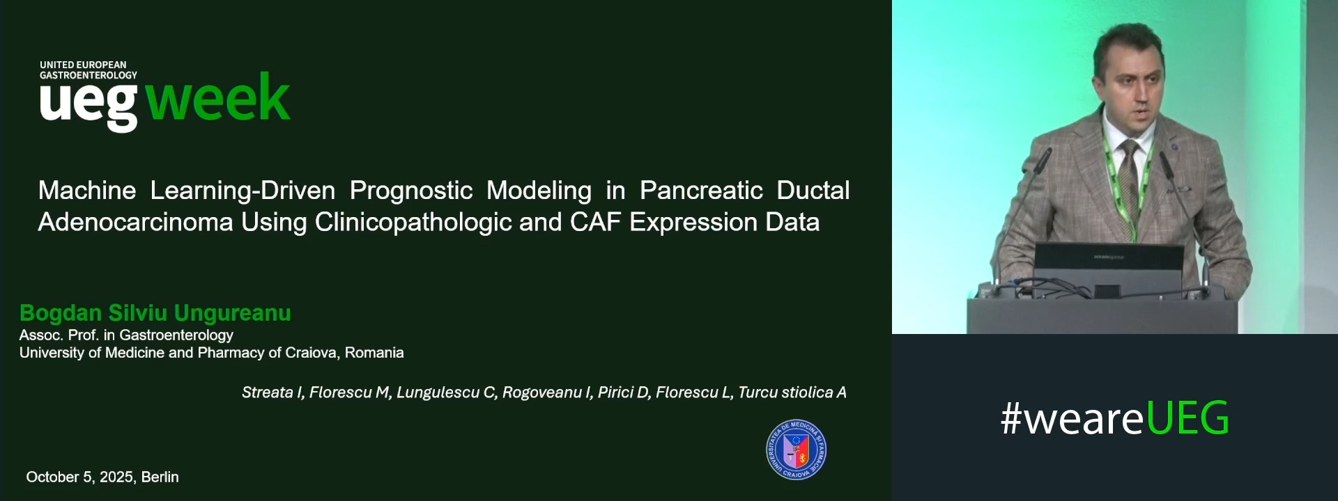
MACHINE LEARNING-DRIVEN PROGNOSTIC MODELING IN PANCREATIC DUCTAL ADENOCARCINOMA USING CLINICOPATHOLOGIC AND CAF EXPRESSION DATA
Bogdan Ungureanu 1, Ioana Streata 2, Mirela Florescu 2, Cristian Lungulescu 2, ION ROGOVEANU 2, Daniel Pirici 2, Lucian Florescu 2, Adina Turcu-Stiolica 2
1 University of Medicine and Pharmacy of Craiova, Craiova, Romania
2 University of Medicine and Pharmacy of Craiova Romania, Craiova, Romania
Event
Topics
Submission format
Citation
Published
Log in to access this content.
Free for all myUEG account holders. Your access level is set automatically based on your occupation. Medical professionals get full access to all content. If you are a non-medical user, you can only access UEG Week content from congresses you attended.
Not sure what you can access? Learn more about account types.
MODELING PRESSURE-TRIGGERED PANCREATIC INFLAMMATION USING HUMAN POLARITY-SWITCHED ORGANOIDS
Aletta Kata Kiss 1, Arpad Varga 1, György Lázár 2, Gergely Groma 3, Ferenc Kagan 4, Petra Pallagi 1, József Maléth 1
1 University of Szeged, Szeged, Hungary|||University of Szeged, Szeged, Hungary|||University of Szeged, Szeged, Hungary
2 University of Szeged, Szeged, Hungary
3 University of Szeged, Szeged, Hungary|||University of Szeged, Szeged, Hungary
4 Max Planck Institute for Multidisciplinary Sciences, Göttingen, Germany
Conference
Topics
Submission format
Session
Citation
Published
Log in to access this content.
Free for all myUEG account holders. Your access level is set automatically based on your occupation. Medical professionals get full access to all content. If you are a non-medical user, you can only access UEG Week content from congresses you attended.
Not sure what you can access? Learn more about account types.
THE ROLE AND UPTAKE OF DRUG AND ALCOHOL SERVICES AMONG PATIENTS WITH ALCOHOL-ASSOCIATED ACUTE PANCREATITIS ADMITTED TO A TERTIARY HEALTH SERVICE
Cindy Ho 1, Alexander Mitropoulos 1, Jeremy Tse 1, Bowen Xia 1, Blake McLeod 1, Olivia Slifirski 1, David Jacka 1, Simon Hew 1
1 Monash Health, Melbourne, Australia
Conference
Topics
Submission format
Session
Citation
Published
Log in to access this content.
Free for all myUEG account holders. Your access level is set automatically based on your occupation. Medical professionals get full access to all content. If you are a non-medical user, you can only access UEG Week content from congresses you attended.
Not sure what you can access? Learn more about account types.
COMPACT PANCREATIC ACINI-SPECIFIC ENHANCER-PROMOTER FOR PANCREATIC GENE THERAPY
1 Changhai Hospital, Shanghai, China
Conference
Topics
Submission format
Session
Citation
Published
Log in to access this content.
Free for all myUEG account holders. Your access level is set automatically based on your occupation. Medical professionals get full access to all content. If you are a non-medical user, you can only access UEG Week content from congresses you attended.
Not sure what you can access? Learn more about account types.
IMPROVING POST-TEST PROBABILITY FOR DIAGNOSIS OF PANCREATIC DUCTAL ADENOCARCINOMA USING A MULTIMODAL ENDOSCOPIC ULTRASOUND APPROACH: A SINGLE CENTER RETROSPECTIVE STUDY INTEGRATING CONTRAST-ENHANCED ENDOSCOPIC ULTRASOUND, STRAIN RATIO ELASTOGRAPHY AND DET
Riccardo Di Pangrazio 1, Leila Babaiyan 1, Matteo Marasco 1, Francesco Panzuto 1, Marianna Signoretti 1
1 Sapienza University, Rome, Italy
Conference
Topics
Submission format
Session
Citation
Published
Log in to access this content.
Free for all myUEG account holders. Your access level is set automatically based on your occupation. Medical professionals get full access to all content. If you are a non-medical user, you can only access UEG Week content from congresses you attended.
Not sure what you can access? Learn more about account types.
THE PROGNOSTIC ROLE OF SYSTEMIC INFLAMMATORY RESPONSE SYNDROME IN ACUTE PANCREATITIS
Teresa Tripiana Iglesias 1, Francisco Valverde-López 1, Ana Lancho Munoz 1, Raúl Mendoza-Rodriguez 1, Eduardo Redondo-Cerezo 2
1 Virgen de las Nieves University Hospital, Granada, Spain
2 Hospital Universitario Virgen de las Nieves, Granada, Spain
Conference
Topics
Submission format
Session