UEG's educational platform for gastroenterology education bringing recordings, courses, guidelines, and more together in one place.
Most popular content
Rate for better recommendations!
Your feedback helps us show you more of what you like and less of what you dislike!
Log in to access this content.
Free for all myUEG account holders. Your access level is set automatically based on your occupation. Medical professionals get full access to all content. If you are a non-medical user, you can only access UEG Week content from congresses you attended.
Not sure what you can access? Learn more about account types.
Timely action in UC: Achieving mucosal healing to optimize patient outcomes (AbbVie) (Complete Session)
1 Department of Medicine 1, Erlangen University Hospital, Friedrich-Alexander-Universität Erlangen-Nürnberg, Erlangen, Germany
2 General University Hospital of Alicante, Spain, None, Spain
3 Inflammatory Bowel Disease Clinic, Calgary, Alberta, Canada
Publisher
Event
Citation
Published
Log in to access this content.
Free for all myUEG account holders. Your access level is set automatically based on your occupation. Medical professionals get full access to all content. If you are a non-medical user, you can only access UEG Week content from congresses you attended.
Not sure what you can access? Learn more about account types.
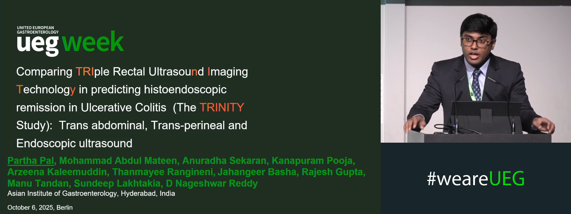
COMPARING TRIPLE RECTAL ULTRASOUND IMAGING TECHNOLOGY IN PREDICTING RECTAL HISTOENDOSCOPIC REMISSION IN ULCERATIVE COLITIS (THE TRINITY STUDY): TRANS ABDOMINAL, TRANS-PERINEAL AND ENDOSCOPIC ULTRASOUND (NCT06717906)
Partha Pal 1, Mohammad Abdul Mateen 1, Anuradha Sekaran 1, Kanapuram Pooja 1, Arzeena Kaleemuddin 1, Thanmayee Rangineni 1, Jahangeer Basha 1, Rajesh Gupta 1, Manu Tandan 1, Sundeep Lakhtakia 1, D. Nageshwar Reddy 1
1 Asian Institute of Gastroenterology, Hyderabad, India
Event
Topics
Submission format
Session
Citation
Published
Log in to access this content.
Free for all myUEG account holders. Your access level is set automatically based on your occupation. Medical professionals get full access to all content. If you are a non-medical user, you can only access UEG Week content from congresses you attended.
Not sure what you can access? Learn more about account types.
Gastric organoids: Insights in mucosal homeostasis and defence against infection
1 Ludwig Cancer Research, Oxford, United Kingdom
Event
Session
Published
Log in to access this content.
Free for all myUEG account holders. Your access level is set automatically based on your occupation. Medical professionals get full access to all content. If you are a non-medical user, you can only access UEG Week content from congresses you attended.
Not sure what you can access? Learn more about account types.
Monitoring ulcerative colitis
1 None, Sde Hemed, Israel
Event
Topics
Published
Log in to access this content.
Free for all myUEG account holders. Your access level is set automatically based on your occupation. Medical professionals get full access to all content. If you are a non-medical user, you can only access UEG Week content from congresses you attended.
Not sure what you can access? Learn more about account types.
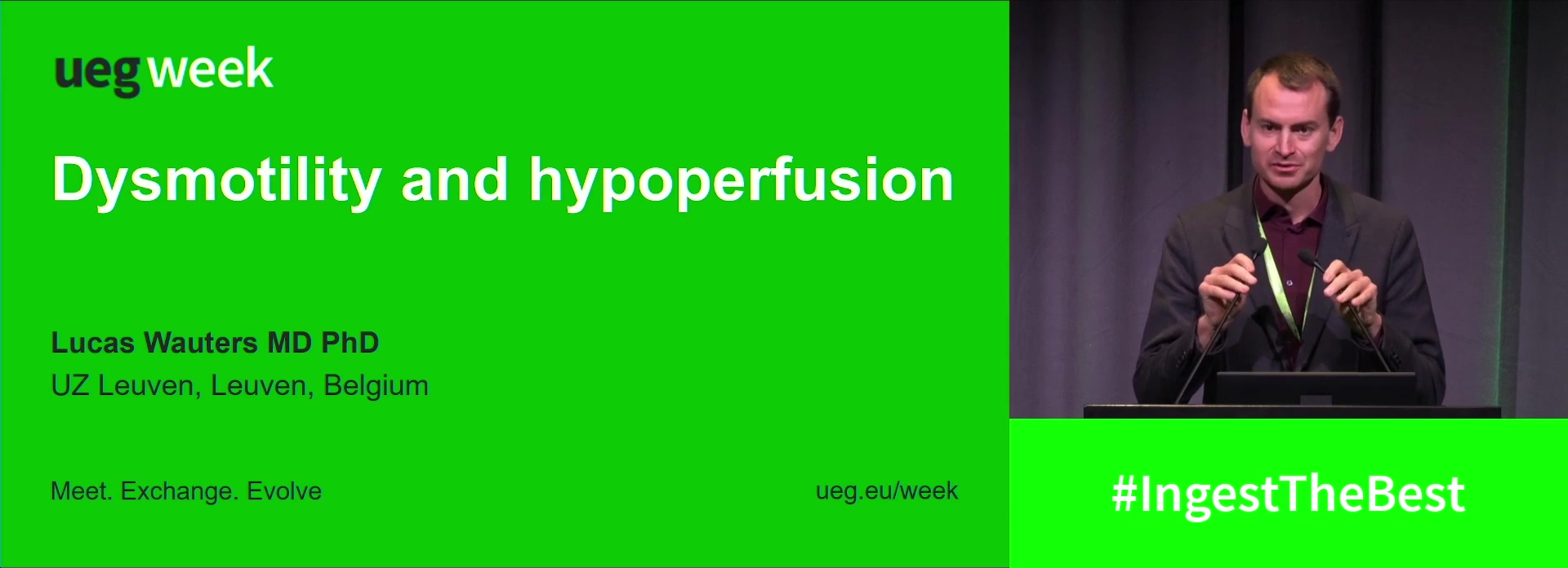
Dysmotility and hypoperfusion
1 University Hospitals Leuven, Leuven, Belgium
Event
Published
Log in to access this content.
Free for all myUEG account holders. Your access level is set automatically based on your occupation. Medical professionals get full access to all content. If you are a non-medical user, you can only access UEG Week content from congresses you attended.
Not sure what you can access? Learn more about account types.
A series of unfortunate events: IBD, flares and CDI (Tillotts Pharma AG) (Complete Session)
1 Beaumont Hospital, Dublin, Ireland, Dublin, Ireland
2 Western University, London, Canada
Event
Citation
Published
Log in to access this content.
Free for all myUEG account holders. Your access level is set automatically based on your occupation. Medical professionals get full access to all content. If you are a non-medical user, you can only access UEG Week content from congresses you attended.
Not sure what you can access? Learn more about account types.
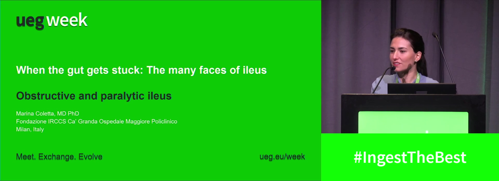
Obstructive and paralytic ileus
1 Università degli Studi di Milano Dept. of Gastroenterology, milan, Italy