UEG's educational platform for gastroenterology education bringing recordings, courses, guidelines, and more together in one place.
Most popular content
Rate for better recommendations!
Your feedback helps us show you more of what you like and less of what you dislike!
Log in to access this content.
Free for all myUEG account holders. Your access level is set automatically based on your occupation. Medical professionals get full access to all content. If you are a non-medical user, you can only access UEG Week content from congresses you attended.
Not sure what you can access? Learn more about account types.

MICROSCOPIC COLITIS: A COMPARATIVE STUDY OF CLINICAL SYMPTOMS AND HISTOPATHOLOGICAL FEATURES
Chaimae Haddad Hachimi 1, Mariam Konso 2, Chaimae Hiji 1, Hayat Ennoufous Krami 3, Mouna Tamzaourte 1
1 Mohamed V Military Training Hospital, Rabat, Morocco
2 Faculty of Medicine and Pharmacy of Rabat, Rabat, Morocco
3 Private Practice, Rabat, Morocco
Conference
Topics
Submission format
Session
Citation
Published
Log in to access this content.
Free for all myUEG account holders. Your access level is set automatically based on your occupation. Medical professionals get full access to all content. If you are a non-medical user, you can only access UEG Week content from congresses you attended.
Not sure what you can access? Learn more about account types.
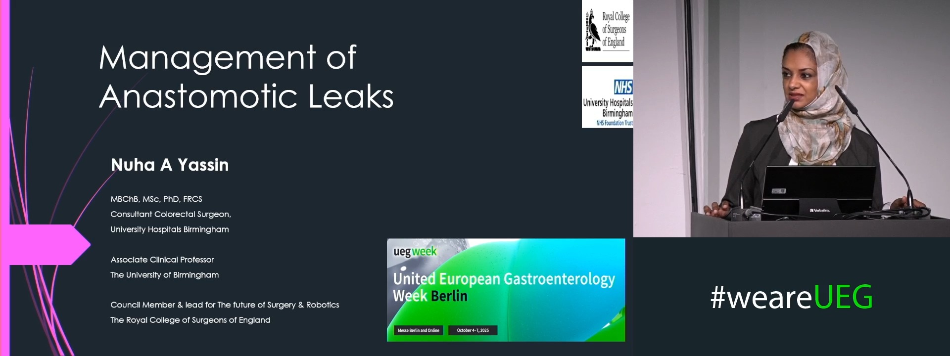
Anastomotic leakage in the gut
1 St Mark'S Hospital, London, London, United Kingdom
Event
Topics
Citation
Published
Log in to access this content.
Free for all myUEG account holders. Your access level is set automatically based on your occupation. Medical professionals get full access to all content. If you are a non-medical user, you can only access UEG Week content from congresses you attended.
Not sure what you can access? Learn more about account types.
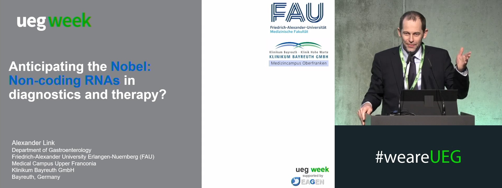
Anticipating the Nobel: Non-coding RNAs in diagnostics and therapy
1 Otto-von-Guericke University of Magdeburg, Magdeburg, Germany
Event
Citation
Published
Log in to access this content.
Free for all myUEG account holders. Your access level is set automatically based on your occupation. Medical professionals get full access to all content. If you are a non-medical user, you can only access UEG Week content from congresses you attended.
Not sure what you can access? Learn more about account types.
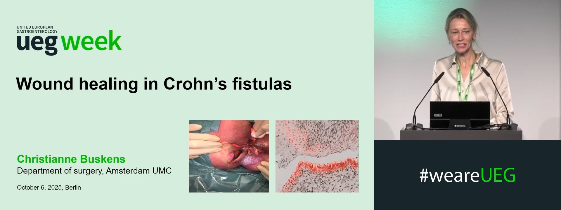
Wound healing in fistula and surgical wounds
1 Academic Medical Center, Amsterdam, Netherlands
Event
Topics
Citation
Published
Log in to access this content.
Free for all myUEG account holders. Your access level is set automatically based on your occupation. Medical professionals get full access to all content. If you are a non-medical user, you can only access UEG Week content from congresses you attended.
Not sure what you can access? Learn more about account types.
Nurse Programme (Complete Session)
Event
Citation
Published
Log in to access this content.
Free for all myUEG account holders. Your access level is set automatically based on your occupation. Medical professionals get full access to all content. If you are a non-medical user, you can only access UEG Week content from congresses you attended.
Not sure what you can access? Learn more about account types.
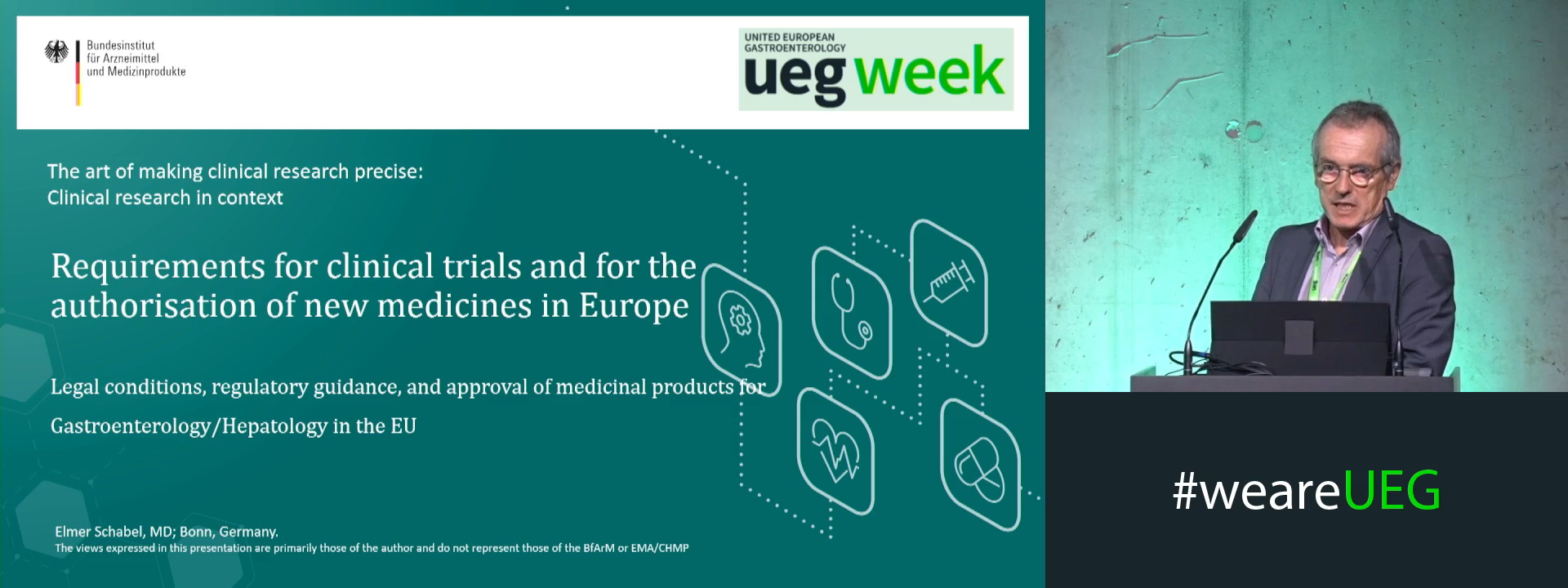
Requirements for clinical trials and for the authorisation of new medicines in Europe
1 Bundesinstitut für Arzneimittel und Medizinprodukte Abt. 2/FG Gastroenterologie, Bonn, Germany
Event
Citation
Published
Log in to access this content.
Free for all myUEG account holders. Your access level is set automatically based on your occupation. Medical professionals get full access to all content. If you are a non-medical user, you can only access UEG Week content from congresses you attended.
Not sure what you can access? Learn more about account types.
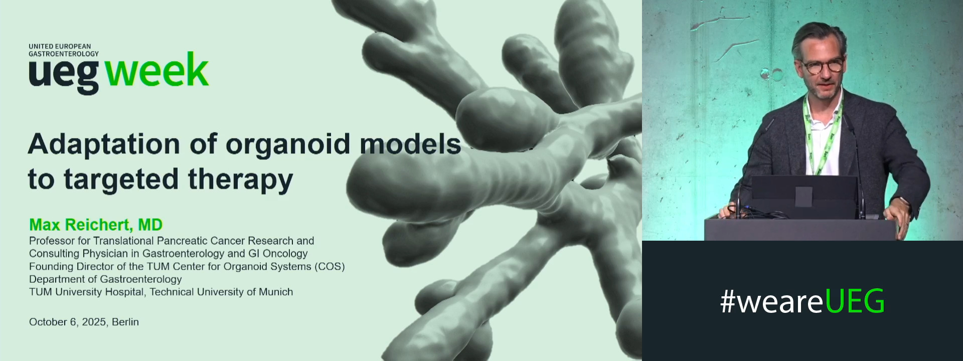
Adaptation of organoid models to targeted therapy
1 TUM Klinikum Rechts der Isar, Munich, Germany
Event
Session