UEG's content platform for gastroenterology education bringing recordings, courses, guidelines, and more together in one place.
Trending on Gutflix
Rate for better recommendations!
Your feedback helps us show you more of what you like and less of what you dislike!
Log in to continue.
This content is part of Gutflix. Log in with your myUEG account, or create one free, to watch it.
Not sure what you can access? Learn more about account types.
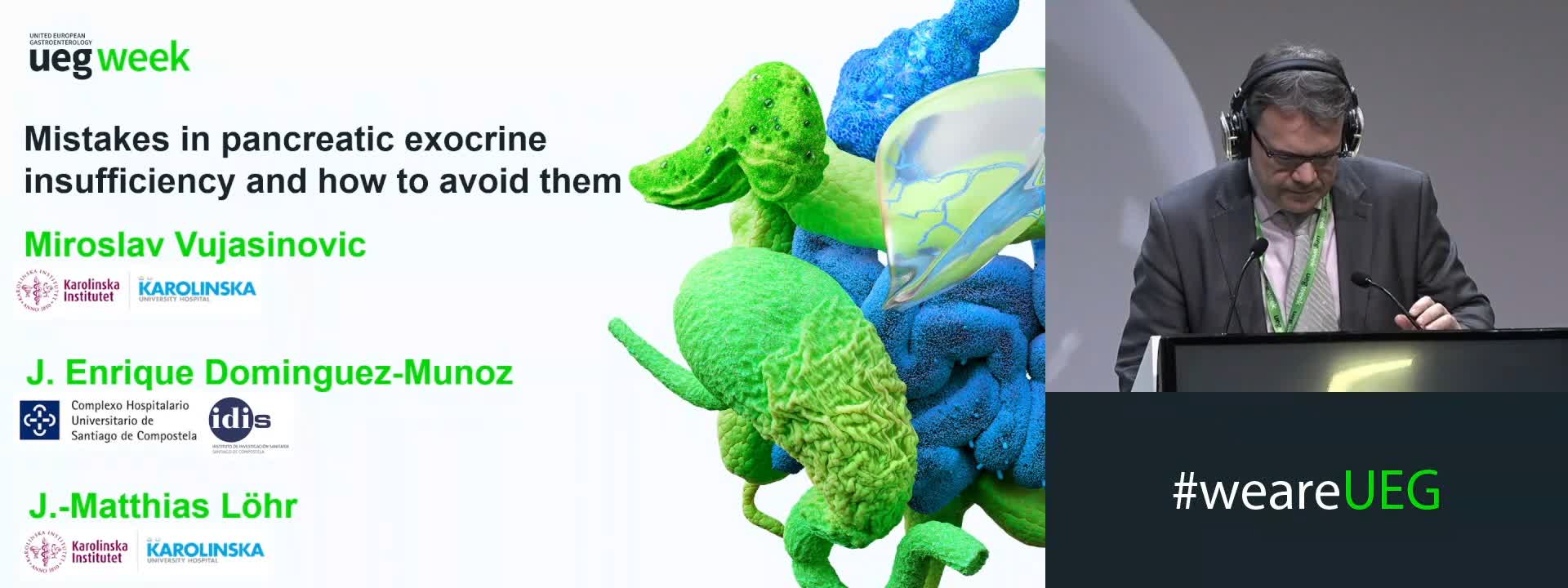
Pancreatic exocrine insufficiency
Affiliations
1 Karolinska University Hospital, Stockholm, Sweden

Log in to continue.
This content is part of Gutflix. Log in with your myUEG account, or create one free, to watch it.
Not sure what you can access? Learn more about account types.
A fusion-based deep-learning algorithm predicting PDAC metastasis from primary tumour
Affiliations
1 Spanish Cancer Research Cancer Center, Madrid - Genetic and Molecular Epidemiology Group, Spanish Ca, Madrid, Spain
Log in to continue.
This content is part of Gutflix. Log in with your myUEG account, or create one free, to watch it.
Not sure what you can access? Learn more about account types.
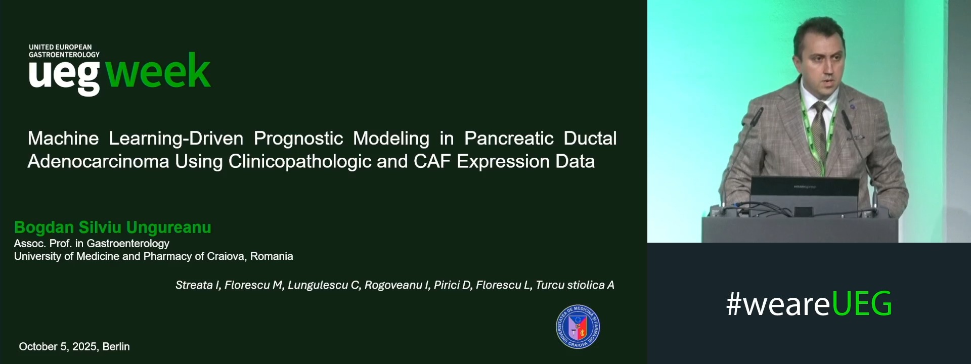
MACHINE LEARNING-DRIVEN PROGNOSTIC MODELING IN PANCREATIC DUCTAL ADENOCARCINOMA USING CLINICOPATHOLOGIC AND CAF EXPRESSION DATA
Bogdan Ungureanu 1, Ioana Streata 2, Mirela Florescu 2, Cristian Lungulescu 2, ION ROGOVEANU 2, Daniel Pirici 2, Lucian Florescu 2, Adina Turcu-Stiolica 2
Affiliations
1 University of Medicine and Pharmacy of Craiova, Craiova, Romania
2 University of Medicine and Pharmacy of Craiova Romania, Craiova, Romania