UEG's educational platform for gastroenterology education bringing recordings, courses, guidelines, and more together in one place.
Most popular content
Rate for better recommendations!
Your feedback helps us show you more of what you like and less of what you dislike!
Log in to access this content.
Free for all myUEG account holders. Your access level is set automatically based on your occupation. Medical professionals get full access to all content. If you are a non-medical user, you can only access UEG Week content from congresses you attended.
Not sure what you can access? Learn more about account types.

IMPACT OF PROBIOTICS ON GUT MICROBIOTA AND METABOLOME DURING HIGH-DOSE DUAL THERAPY WITH VONOPRAZAN AND AMOXICILLIN AS HELICOBACTER PYLORI RESCUE THERAPY: A PROSPECTIVE, MULTICENTER, RANDOMIZED CONTROLLED STUDY
1 Zhongshan Hospital (Xiamen), Fudan University, Xiamen, China
2 Third Hospital of Xiamen, Xiamen, China
3 Second Affiliated Hospital of Xiamen Medical College, Xiamen, China
4 Zhongshan Hospital Fudan University, Shanghai, China
Conference
Topics
Submission format
Citation
Published
Log in to access this content.
Free for all myUEG account holders. Your access level is set automatically based on your occupation. Medical professionals get full access to all content. If you are a non-medical user, you can only access UEG Week content from congresses you attended.
Not sure what you can access? Learn more about account types.
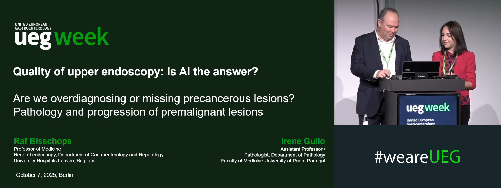
Premalignant gastric lesions: Pathology, progressions, and challenges in diagnosis
1 Katholieke Universiteit Leuven, Leuven, Belgium
2 Faculty of Medicine of the University of Porto, Porto, Portugal
Event
Citation
Published
Log in to access this content.
Free for all myUEG account holders. Your access level is set automatically based on your occupation. Medical professionals get full access to all content. If you are a non-medical user, you can only access UEG Week content from congresses you attended.
Not sure what you can access? Learn more about account types.
HISTOPATHOLOGICAL CORRELATIONS OF TISSUE SOUND SPEED IN HEPATOCELLULAR CARCINOMA: A QUANTITATIVE ASSESSMENT USING ACOUSTIC MICROSCOPY
1 Graduate School of Medicine, Tohoku University, Sendai, Japan|||Saitama Cooperative Hospital, Kawaguchi, Japan
2 Graduate School of Medicine, Tohoku University, Sendai, Japan
3 Honda Electronics, Co., Ltd., Toyohashi, Japan
4 Saitama Cooperative Hospital, Kawaguchi, Japan
Conference
Topics
Submission format
Session
Citation
Published
Log in to access this content.
Free for all myUEG account holders. Your access level is set automatically based on your occupation. Medical professionals get full access to all content. If you are a non-medical user, you can only access UEG Week content from congresses you attended.
Not sure what you can access? Learn more about account types.
PAPILLARY LARGE BALLOON DILATATION VERSUS ENDOSCOPIC COMPLETE SPHINCTEROTOMY IN PATIENTS WITH LARGE COMMON BILE DUCT STONE(S) AND PERIAMPULLARY DIVERTICULUM
Ibrahim M. Ibrahim 1, Tarek I. Zaher 1, Soha E. Khorshed 1, mahmoud sheded 2, Tarek Mostafa Ibrahim Mohammed 2, Ahmed Atya 1, Mohamed Hassan Emara Elzanan 3, Mohammed Ahmed Hasuna Hemeda 1
1 Zagazig University, Zagazig, Egypt
2 Suez University, Suez, Egypt
3 Kafrelsheikh University, Kafr Elshiekh, Egypt|||Alyousif Hospital, Alkhobar, Saudi Arabia
Conference
Topics
Submission format
Citation
Published
Log in to access this content.
Free for all myUEG account holders. Your access level is set automatically based on your occupation. Medical professionals get full access to all content. If you are a non-medical user, you can only access UEG Week content from congresses you attended.
Not sure what you can access? Learn more about account types.
ROLE OF ENDOSCOPIC BILIARY STENTING IN ACCESS TO CHEMOTHERAPY IN PATIENTS WITH UNRESECTABLE MALIGNANT OBSTRUCTION
1 Medical University of Silesia, Katowice, Poland
2 Univeristy of Silesia, Katowice, Poland
Conference
Topics
Submission format
Session
Citation
Published
Log in to access this content.
Free for all myUEG account holders. Your access level is set automatically based on your occupation. Medical professionals get full access to all content. If you are a non-medical user, you can only access UEG Week content from congresses you attended.
Not sure what you can access? Learn more about account types.
FECAL MICROBIOTA TRANSPLANTATION FOR CHILDREN WITH AUTISM SPECTRUM DISORDER AND THE CORE FUNCTIONAL STRAIN IDENTIFICATION: A COHORT STUDY
Dongling Dai 1, Zhongsheng Zhu 2, Yigui Zou 1, Ziyuan Li 1, Boxian Lin 1, Mingjing Luo 3, Haokui Zhou 3
1 Shenzhen Children's Hospital, Shenzhen, China
2 Shenzhen Children’s Hospital, Shenzhen, China
3 Shenzhen Institute of Advanced Technology, Chinese Academy of Sciences, Shenzhen, China
Conference
Topics
Submission format
Session
Citation
Published

Log in to access this content.
Free for all myUEG account holders. Your access level is set automatically based on your occupation. Medical professionals get full access to all content. If you are a non-medical user, you can only access UEG Week content from congresses you attended.
Not sure what you can access? Learn more about account types.
IMPACT OF FIRST AND FURTHER DECOMPENSATION IN METABOLIC-DYSFUNCTION ASSOCIATED COMPENSATED ADVANCED CHRONIC LIVER DISEASE
Grazia Pennisi 1, Gabriele Di Maria 2, Vincent Wai Sun Wong 3, Victor De Ledinghen 4, Giada Sebastiani 5, Mauro Vigano 6, Anna Francanzani 7, Luca Miele 8, Elisabetta Bugianesi 9, Mattias Ekstedt 10, Roberta D'Ambrosio 11, Federico Ravaioli 12, Filippo Schepis 13, Fabio Marra 14, Alessio Aghemo 15, Gianluca Svegliati Baroni 16, Marcello Persico 17, Luca Valenti 6, Annalisa Berzigotti 18, Jacob George 19, Angelo Armandi 20, Patrik Nasr 10, Stergios Kechagias 10, Antonio Liguori 21, Dario Saltini 13, YULY PAULIN MENDOZA JAIMES 22, Vincenza Calvaruso 2, Marco Enea 2, Huapeng Lin 23, Giuseppe Infantino 2, Mario Masarone 17, Nicola Pugliese 24, Adele Tulone 2, Vito Di Marco 2, Calogero Camma 25, Salvatore Petta 26
1 Policlinico Paolo Giaccone, Palermo, Italy
2 University of Palermo, Palermo, Italy
3 University of Hong Kong, Hong Kong, China
4 Bordeaux University Hospital, Pessac, France
5 McGill University Health Centre, Montreal, Canada
6 University of Milan, Milan, Italy
7 Università degli Studi di Milano, Fondazione Ospedale Policlinico Ca Granda IRCCS, Milan, Italy
8 Fondazione Policlinico Gemelli IRCCS, Università Cattolica S. Cuore, Rome, Italy
9 AOU Città della Salute e della Scienza University of Torino, Torino, Italy, Torino, Italy
10 Linköping University, Linköping, Sweden
11 Ospedale Maggiore Policlinico, Università degli Studi di Milano, Milan, Italy
12 University of Bologna, Bologna, Italy
13 University of Modena, Modena, Italy
14 University of Florence, Florence, Italy
15 Humanitas University, Pieve Emanuele, Italy
16 Università Politecnica delle Marche, Ancona, Italy
17 University of Salerno, Salerno, Italy
18 Inselspital, University Hospital of Berne, Berne, Switzerland
19 University of Sydney, Sidney, Italy
20 University of Turin, Torino, Italy
21 Catholic University Of Sacred Heart, Rome, Rome, Italy
22 Inselspital, DBMR, university of Berm, Bern, Switzerland
23 The Chinese University of Hong Kong, Hong Kong, Italy
24 Istituto Clinico Humanitas IRCCS, Rozzano, Italy
25 UOC Gastroenterologia Ed Epatologia, Palermo, Italy
26 University of Palermo Italy 48.37, Palermo, Italy