UEG's educational platform for gastroenterology education bringing recordings, courses, guidelines, and more together in one place.
Most popular content
Rate for better recommendations!
Your feedback helps us show you more of what you like and less of what you dislike!
Log in to access this content.
Free for all myUEG account holders. Your access level is set automatically based on your occupation. Medical professionals get full access to all content. If you are a non-medical user, you can only access UEG Week content from congresses you attended.
Not sure what you can access? Learn more about account types.
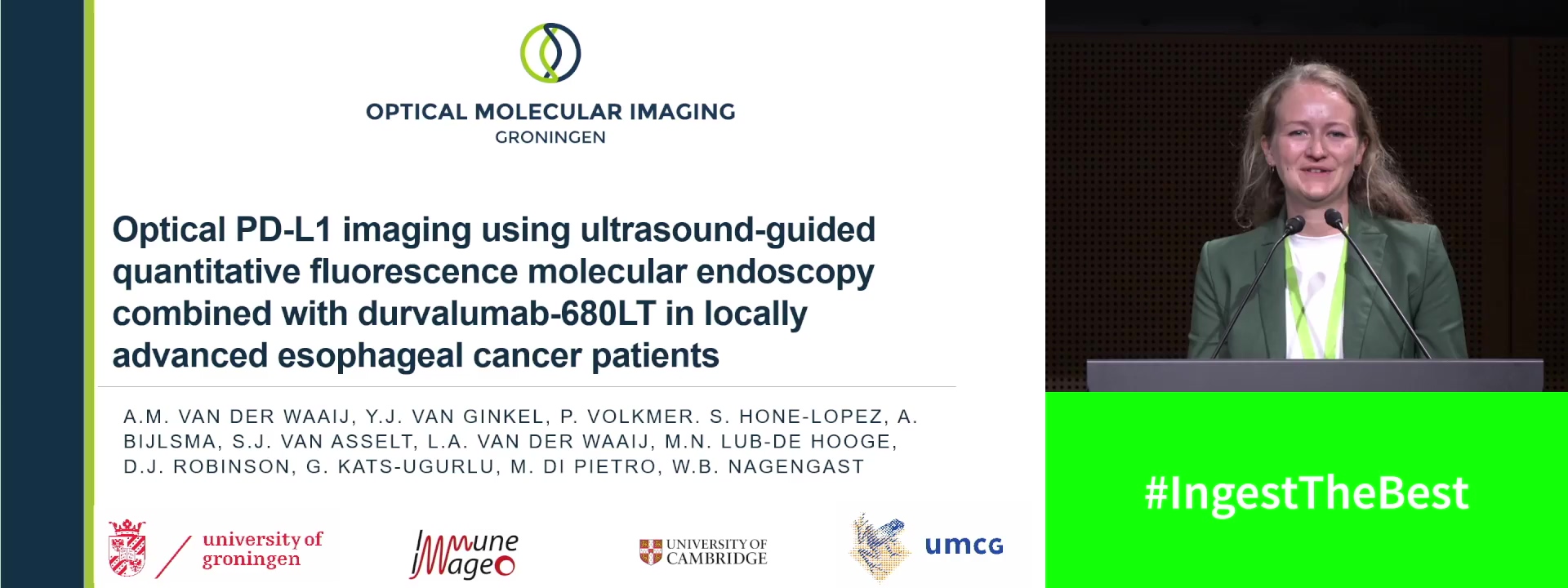
OPTICAL PD-L1 IMAGING USING ULTRASOUND-GUIDED QUANTITATIVE FLUORESCENCE MOLECULAR ENDOSCOPY COMBINED WITH DURVALUMAB-680LT IN LOCALLY ADVANCED ESOPHAGEAL CANCER PATIENTS
Anne M. van der Waaij 1, Ymke van Ginkel 1, Sara Hone-Lopez 1, Rina Bijlsma 2, Sophie J. van Asselt 2, Laurens A. van der Waaij 2, Marjolijn N. Lub-de Hooge 1, Dominic James Robinson 3, Gursah Kats-Ugurlu 1, Massimiliano di Pietro 4, Wouter B. Nagengast 1
1 University Medical Center Groningen, Groningen, Netherlands
2 Martini Hospital, Groningen, Netherlands
3 Center for Optic Diagnostics and Therapy, Rotterdam, Netherlands
4 University of Cambridge, Cambridge, United Kingdom
Event
Submission format
Citation
Published
Log in to access this content.
Free for all myUEG account holders. Your access level is set automatically based on your occupation. Medical professionals get full access to all content. If you are a non-medical user, you can only access UEG Week content from congresses you attended.
Not sure what you can access? Learn more about account types.
Log in to access this content.
Free for all myUEG account holders. Your access level is set automatically based on your occupation. Medical professionals get full access to all content. If you are a non-medical user, you can only access UEG Week content from congresses you attended.
Not sure what you can access? Learn more about account types.
The future of immunotherapy → are surgeons obsolete soon? with Jeroen Dekervel
Topics
Published
Log in to access this content.
Free for all myUEG account holders. Your access level is set automatically based on your occupation. Medical professionals get full access to all content. If you are a non-medical user, you can only access UEG Week content from congresses you attended.
Not sure what you can access? Learn more about account types.
Log in to access this content.
Free for all myUEG account holders. Your access level is set automatically based on your occupation. Medical professionals get full access to all content. If you are a non-medical user, you can only access UEG Week content from congresses you attended.
Not sure what you can access? Learn more about account types.
The Future of Faecal Microbiota Transplantation (FMT) with Maria Vehreschild
Topics
Published
Log in to access this content.
Free for all myUEG account holders. Your access level is set automatically based on your occupation. Medical professionals get full access to all content. If you are a non-medical user, you can only access UEG Week content from congresses you attended.
Not sure what you can access? Learn more about account types.
From Vienna to Berlin: What inspired us last year at UEG Week
Published
Log in to access this content.
Free for all myUEG account holders. Your access level is set automatically based on your occupation. Medical professionals get full access to all content. If you are a non-medical user, you can only access UEG Week content from congresses you attended.
Not sure what you can access? Learn more about account types.