UEG Week Recordings UEG Week Posters Online courses Guidelines Mistakes in... Podcasts Webinars
Visit ueg.eu Create myUEG account Log In

Filters:

UEG Mistakes In Articles
Share via Email Share on Facebook Share on X Share on LinkedIn Share on Bluesky

Log in to access this content.

Free for all myUEG account holders. Your access level is set automatically based on your occupation. Medical professionals get full access to all content. If you are a non-medical user, you can only access UEG Week content from congresses you attended.

Log In Create a free account

Not sure what you can access? Learn more about account types.

Dealing with upper gastrointestinal (UGI) bleeding is complex and challenging, especially since distinguishing patients with significant bleeding from those with other causes of illness and hypotension can be difficult. Despite these diagnostic challenges and the increasing age and comorbidities of patients, the mortality rate from UGI bleeding has remained stable over the past 30 years. Mortality rates vary in published studies, often due to the inclusion of healthier patients without significant bleeding. The best way to assess outcomes is by focusing on patients with bleeding ulcers and varices, with a noted 30-day mortality rate of 22% in such cases over a five-year period in Leeds. The standard of care for emergency GI bleeding involves offering an emergency gastroscopy within 24 hours, although this benchmark is not always met in the UK. Early endoscopy is known to be safe, reduces hospital stay length, and decreases the need for emergency surgery, though there is no strong evidence that it directly saves lives. This may be because many studies include patients without significant bleeding. Drawing on years of clinical experience, this discussion will focus on common mistakes made in managing UGI bleeding.

Mistakes in upper gastrointestinal bleeding and how to avoid them

Mistakes in upper gastrointestinal bleeding and how to avoid them

Bjorn Rembacken

Topics

Digestive Oncology Endoscopy Stomach & H. Pylori

Citation

Rembacken BJ. Mistakes in upper gastrointestinal bleeding and how to avoid them. UEG Education 2016: 16: 15–19.

Published

2016
UEG Podcast Episode
Journal Podcast
Share via Email Share on Facebook Share on X Share on LinkedIn Share on Bluesky

Log in to access this content.

Free for all myUEG account holders. Your access level is set automatically based on your occupation. Medical professionals get full access to all content. If you are a non-medical user, you can only access UEG Week content from congresses you attended.

Log In Create a free account

Not sure what you can access? Learn more about account types.

Episode 6: UEG Journal October Spotlight

Mohsan Subhani, Maria Manuela Estevinho

Topics

Endoscopy Hepatobiliary IBD Pancreas

Published

2025
UEG Mistakes In Articles
Share via Email Share on Facebook Share on X Share on LinkedIn Share on Bluesky

Log in to access this content.

Free for all myUEG account holders. Your access level is set automatically based on your occupation. Medical professionals get full access to all content. If you are a non-medical user, you can only access UEG Week content from congresses you attended.

Log In Create a free account

Not sure what you can access? Learn more about account types.

Hepatitis C virus (HCV) infection remains an important global health concern. It is estimated that there are approximately 50 million people infected with HCV globally, with around 1 million new infections each year and about 242,000 deaths annually attributed to HCV-related complications. Most acute HCV infections (55–85%) become chronic due to the virus’s effective evasion strategies, with spontaneous clearance being rare once chronicity is established. This condition often progresses silently, with many individuals unaware of their infection until advanced liver damage has occurred. If left untreated, HCV can lead to severe complications, including liver cirrhosis and hepatocellular carcinoma (HCC). HCV transmission occurs mainly through percutaneous exposure to infected blood. HCV can also spread from mother to infant (vertical transmission) and, less frequently, via sexual contact.1,2 In recent years, the introduction of oral direct-acting antivirals (DAAs), with remarkable safety and effectiveness profiles, has led to a sustained virological response (SVR) in virtually all (>97%) HCV-infected patients, regardless of HCV genotype or disease stage. However, significant barriers remain, such as issues with diagnosis, access to treatment and awareness of the disease.

Here, we discuss some of the misconceptions in HCV management and provide a practical management approach grounded in evidence and clinical experience.

Mistakes in hepatitis C and how to avoid them

Mistakes in hepatitis C and how to avoid them

Ana Catarina Garcia, Gonçalo Alexandrino

Topics

Hepatobiliary

Citation

Garcia A.C and Alexandrino G. Mistakes in hepatits C and how to avoid them. UEG Education 2025; 25: 14-17.

Published

2025
UEG Mistakes In Articles
Share via Email Share on Facebook Share on X Share on LinkedIn Share on Bluesky

Log in to access this content.

Free for all myUEG account holders. Your access level is set automatically based on your occupation. Medical professionals get full access to all content. If you are a non-medical user, you can only access UEG Week content from congresses you attended.

Log In Create a free account

Not sure what you can access? Learn more about account types.

Long-term enteral nutrition via gastrostomy is a relatively common medical intervention for patients at risk of malnutrition who have an accessible and functioning gastrointestinal tract. There are clear clinical guidelines describing the principles of practice as well as numerous retrospective and non-randomised controlled studies and case series. However, fewer publications impart advice and guidance regarding the management and ‘patient selection’ for these interventions. The following article provides a combination of the author’s views and the evidence base.

Mistakes in gastrostomy insertion and how to avoid them

Mistakes in gastrostomy insertion and how to avoid them

Tom Welbank

Topics

Small Intestine & Nutrition Stomach & H. Pylori

Citation

Welbank T, Mistakes in gastrostomy insertion ingestion and how to avoid them. UEG Education 2024; 24: 8-11.

Published

2024
UEG Standards and Guidelines
Clinical Practice Guideline
Share via Email Share on Facebook Share on X Share on LinkedIn Share on Bluesky

Log in to access this content.

Free for all myUEG account holders. Your access level is set automatically based on your occupation. Medical professionals get full access to all content. If you are a non-medical user, you can only access UEG Week content from congresses you attended.

Log In Create a free account

Not sure what you can access? Learn more about account types.

Main Recommendations

At a population level, the European Society of Gastrointestinal Endoscopy (ESGE), the European Helicobacter and Microbiota Study Group (EHMSG), and the European Society of Pathology (ESP) suggest endoscopic screening for gastric cancer (and precancerous conditions) in high-risk regions (age-standardized rate [ASR] > 20 per 100 000 person-years) every 2 to 3 years or, if cost–effectiveness has been proven, in intermediate risk regions (ASR 10–20 per 100 000 person-years) every 5 years, but not in low-risk regions (ASR < 10).

ESGE/EHMSG/ESP recommend that irrespective of country of origin, individual gastric risk assessment and stratification of precancerous conditions is recommended for first-time gastroscopy.

ESGE/EHMSG/ESP suggest that gastric cancer screening or surveillance in asymptomatic individuals over 80 should be discontinued or not started, and that patients’ comorbidities should be considered when treatment of superficial lesions is planned.

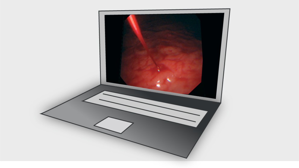
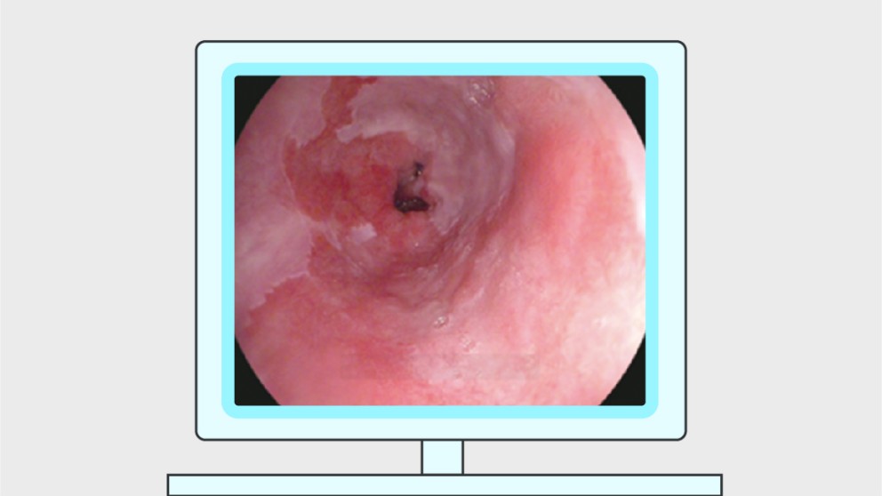
ESGE/EHMSG/ESP recommend that a high quality endoscopy including the use of virtual chromoendoscopy (VCE), after proper training, is performed for screening, diagnosis, and staging of precancerous conditions (atrophy and intestinal metaplasia) and lesions (dysplasia or cancer), as well as after endoscopic therapy. VCE should be used to guide the sampling site for biopsies in the case of suspected neoplastic lesions as well as to guide biopsies for diagnosis and staging of gastric precancerous conditions, with random biopsies to be taken in the absence of endoscopically suspected changes. When there is a suspected early gastric neoplastic lesion, it should be properly described (location, size, Paris classification, vascular and mucosal pattern), photodocumented, and two targeted biopsies taken.

ESGE/EHMSG/ESP do not recommend routine performance of endoscopic ultrasonography (EUS), computed tomography (CT), magnetic resonance imaging (MRI), or positron emission tomography (PET)-CT prior to endoscopic resection unless there are signs of deep submucosal invasion or if the lesion is not considered suitable for endoscopic resection.

ESGE/EHMSG/ESP recommend endoscopic submucosal dissection (ESD) for differentiated gastric lesions clinically staged as dysplastic (low grade and high grade) or as intramucosal carcinoma (of any size if not ulcerated or ≤ 30 mm if ulcerated), with EMR being an alternative for Paris 0-IIa lesions of size ≤ 10 mm with low likelihood of malignancy.

ESGE/EHMSG/ESP suggest that a decision about ESD can be considered for malignant lesions clinically staged as having minimal submucosal invasion if differentiated and ≤ 30 mm; or for malignant lesions clinically staged as intramucosal, undifferentiated and ≤ 20 mm; and in both cases with no ulcerative findings.

ESGE/EHMSG/ESP recommends patient management based on the following histological risk after endoscopic resection:

Curative/very low-risk resection (lymph node metastasis [LNM] risk < 0.5 %–1 %): en bloc R0 resection; dysplastic/pT1a, differentiated lesion, no lymphovascular invasion, independent of size if no ulceration and ≤ 30 mm if ulcerated. No further staging procedure or treatment is recommended.

Curative/low-risk resection (LNM risk < 3 %): en bloc R0 resection; lesion with no lymphovascular invasion and: a) pT1b, invasion ≤ 500 µm, differentiated, size ≤ 30 mm; or b) pT1a, undifferentiated, size ≤ 20 mm and no ulceration. Staging should be completed, and further treatment is generally not necessary, but a multidisciplinary discussion is required.

Local-risk resection (very low risk of LNM but increased risk of local persistence/recurrence): Piecemeal resection or tumor-positive horizontal margin of a lesion otherwise meeting curative/very low-risk criteria (or meeting low-risk criteria provided that there is no submucosal invasive tumor at the resection margin in the case of piecemeal resection or tumor-positive horizontal margin for pT1b lesions [invasion ≤ 500 µm; well-differentiated; size ≤ 30 mm, and VM0]). Endoscopic surveillance/re-treatment is recommended rather than other additional treatment.

High-risk resection (noncurative): Any lesion with any of the following: (a) a positive vertical margin (if carcinoma) or lymphovascular invasion or deep submucosal invasion (> 500 µm from the muscularis mucosae); (b) poorly differentiated lesions if ulceration or size > 20 mm; (c) pT1b differentiated lesions with submucosal invasion ≤ 500 µm with size > 30 mm; or (d) intramucosal ulcerative lesion with size > 30 mm. Complete staging and strong consideration for additional treatments (surgery) in multidisciplinary discussion.

ESGE/EHMSG/ESP suggest the use of validated endoscopic classifications of atrophy (e. g. Kimura–Takemoto) or intestinal metaplasia (e. g. endoscopic grading of gastric intestinal metaplasia [EGGIM]) to endoscopically stage precancerous conditions and stratify the risk for gastric cancer.

ESGE/EHMSG/ESP recommend that biopsies should be taken from at least two topographic sites (2 biopsies from the antrum/incisura and 2 from the corpus, guided by VCE) in two separate, clearly labeled vials. Additional biopsy from the incisura is optional.

ESGE/EHMSG/ESP recommend that patients with extensive endoscopic changes (Kimura C3 + or EGGIM 5 +) or advanced histological stages of atrophic gastritis (severe atrophic changes or intestinal metaplasia, or changes in both antrum and corpus, operative link on gastritis assessment/operative link on gastric intestinal metaplasia [OLGA/OLGIM] III/IV) should be followed up with high quality endoscopy every 3 years, irrespective of the individual’s country of origin.

ESGE/EHMSG/ESP recommend that no surveillance is proposed for patients with mild to moderate atrophy or intestinal metaplasia restricted to the antrum, in the absence of endoscopic signs of extensive lesions or other risk factors (family history, incomplete intestinal metaplasia, persistent H. pylori infection). This group constitutes most individuals found in clinical practice.

ESGE/EHMSG/ESP recommend H. pylori eradication for patients with precancerous conditions and after endoscopic or surgical therapy.

ESGE/EHMSG/ESP recommend that patients should be advised to stop smoking and low-dose daily aspirin use may be considered for the prevention of gastric cancer in selected individuals with high risk for cardiovascular events.

Management of epithelial precancerous conditions and early neoplasia of the stomach (MAPS III): ESGE, EHMSG and ESP Guideline update 2025

Management of epithelial precancerous conditions and early neoplasia of the stomach (MAPS III): ESGE, EHMSG and ESP Guideline update 2025

Mario Dinis-Ribeiro

Publishers

European Society of Pathology logoEuropean Helicobacter and Microbiota Study Group logoEuropean Society of Gastrointestinal Endoscopy logo
European Society of Pathology, European Helicobacter and Microbiota Study Group, European Society of Gastrointestinal Endoscopy

Guideline

Clinical Practice Guideline

Topics

Endoscopy Stomach & H. Pylori

Citation

Endoscopy 2025; 57(05): 504-554

Published

2025
Login to access
UEG Podcast Episode
UEG Podcast
Share via Email Share on Facebook Share on X Share on LinkedIn Share on Bluesky

Log in to access this content.

Free for all myUEG account holders. Your access level is set automatically based on your occupation. Medical professionals get full access to all content. If you are a non-medical user, you can only access UEG Week content from congresses you attended.

Log In Create a free account

Not sure what you can access? Learn more about account types.

Oesophageal cancer with Massimiliano di Pietro (Part 2)

Massimiliano di Pietro, Pradeep Mundre

Topics

Digestive Oncology Endoscopy Oesophagus

Published

2025
UEG Mistakes In Articles
Share via Email Share on Facebook Share on X Share on LinkedIn Share on Bluesky

Log in to access this content.

Free for all myUEG account holders. Your access level is set automatically based on your occupation. Medical professionals get full access to all content. If you are a non-medical user, you can only access UEG Week content from congresses you attended.

Log In Create a free account

Not sure what you can access? Learn more about account types.

Barrett’s oesophagus is the precursor to oesophageal adenocarcinoma, which carries a poor prognosis, and it is likely that all endoscopists and gastroenterologists will encounter Barrett’s oesophagus in their clinical practice. Careful assessment and management of patients who have Barrett’s oesophagus with endoscopic surveillance and endoscopic endotherapy aim to reduce the risk of progression to invasive adenocarcinoma. Advances in endoscopic diagnosis and therapy should, therefore, help to reduce the risk of progression. As with all premalignant conditions and surveillance programmes, careful multidisciplinary management of the patient is important to reduce the risk of causing them to become unduly concerned. Here, we present some mistakes that in our experience are commonly made in the endoscopic diagnosis and management of Barrett’s oesophagus and give advice on how to avoid them. 

Mistakes in the endoscopic diagnosis and management of Barrett’s oesophagus and how to avoid them

Mistakes in the endoscopic diagnosis and management of Barrett’s oesophagus and how to avoid them

Apostolis Papaefthymiou, Cormac Magee, Rehan Jamil Haidry

Topics

Endoscopy Oesophagus

Citation

Haidry RJ and Magee C. Mistakes in the endoscopic diagnosis and management of Barrett’s oesophagus and how to avoid them. UEG Education 2018; 2018: 12–14.

Published

2024

The global reference point for the digestive health community

Platform Publisher

United European Gastroenterology

Wickenburggasse 1 1080 Vienna, Austria

Contact us

support@ueg.eu

ueg.eu

T: +43 1 997 1639

Legal

Terms & Conditions

Imprint

Privacy Policy

Explore

My Bookmarks

My recommendations

My fields of interest

© 2026 United European Gastroenterology

Change fields of interest

These fields are selected based on the interests in your myUEG profile.
Click the item to unselect it. You can select multiple items.