UEG's content platform for gastroenterology education bringing recordings, courses, guidelines, and more together in one place.
Trending on Gutflix
Rate for better recommendations!
Your feedback helps us show you more of what you like and less of what you dislike!
Log in to continue.
This content is part of Gutflix. Log in with your myUEG account, or create one free, to watch it.
Not sure what you can access? Learn more about account types.
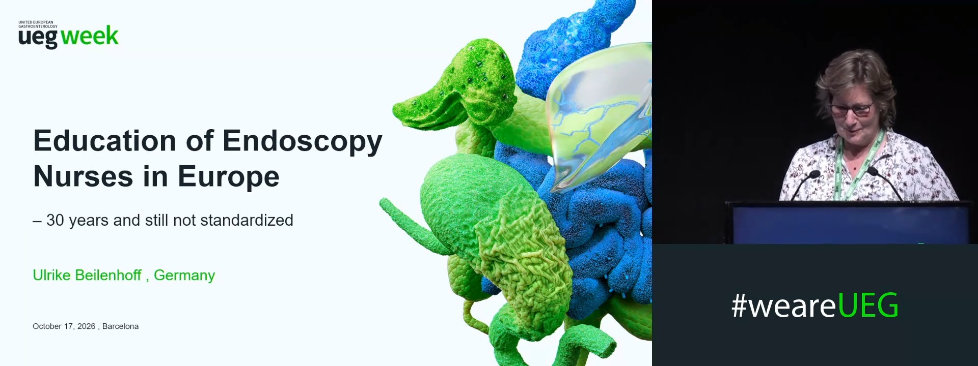
Education of endoscopy nurses in Europe – 30 years and still not standardised
Affiliations
1 ESGENA Scientific Secretary, Ulm, Germany
Log in to continue.
This content is part of Gutflix. Log in with your myUEG account, or create one free, to watch it.
Not sure what you can access? Learn more about account types.
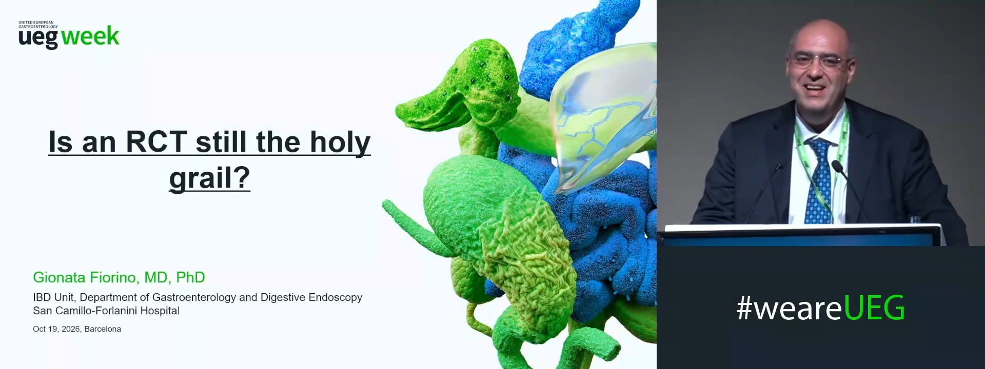
Is an RCT still the holy grail?
Affiliations
1 IRCCS Humanitas, Milan, Italy
Log in to continue.
This content is part of Gutflix. Log in with your myUEG account, or create one free, to watch it.
Not sure what you can access? Learn more about account types.
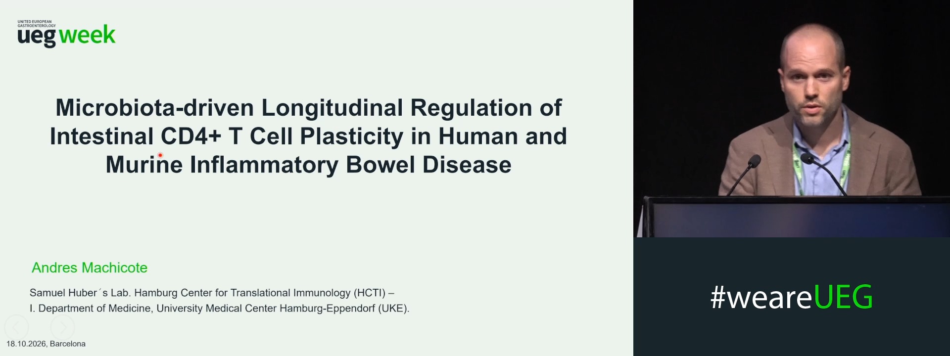
MICROBIOTA-DRIVEN LONGITUDINAL REGULATION OF INTESTINAL CD4+ T CELL PLASTICITY IN HUMAN AND MURINE INFLAMMATORY BOWEL DISEASE
Andres Machicote 1,2, Beibei Liu 1,2, Marie Wende 3, Franziska Stallbaum 1,2, Babett Steglich 1,2, Ban H. Tran 1,2, Sandra Wende 1,2, Saskia Großhauser 1,2, Zehra Oguz-Cöloglu 1, Till Robin Lesker 3, Moritz von Wulffen 1, Penelope Pelczar 2,1, Tanja Bedke 1,2, Mikolaj Nawrocki 1,2, Marius Böttcher 1, Jan Kempski 1, Katharina Moll 1, Thorben W. Fründt 1, Thomas Rösch 1, Till Strowig 3, Nicola Gagliani 4,2, Samuel Huber 1,2
Affiliations
1 Universitätsklinikum Hamburg-Eppendorf (UKE), Hamburg, Germany
2 Hamburg Center for Translational Immunology, Hamburg, Germany
3 Helmholtz-Zentrum für Infektionsforschung, Braunschweig, Germany
4 Institute for Inflammation and Carcinogenesis, Hamburg, Germany
Log in to continue.
This content is part of Gutflix. Log in with your myUEG account, or create one free, to watch it.
Not sure what you can access? Learn more about account types.
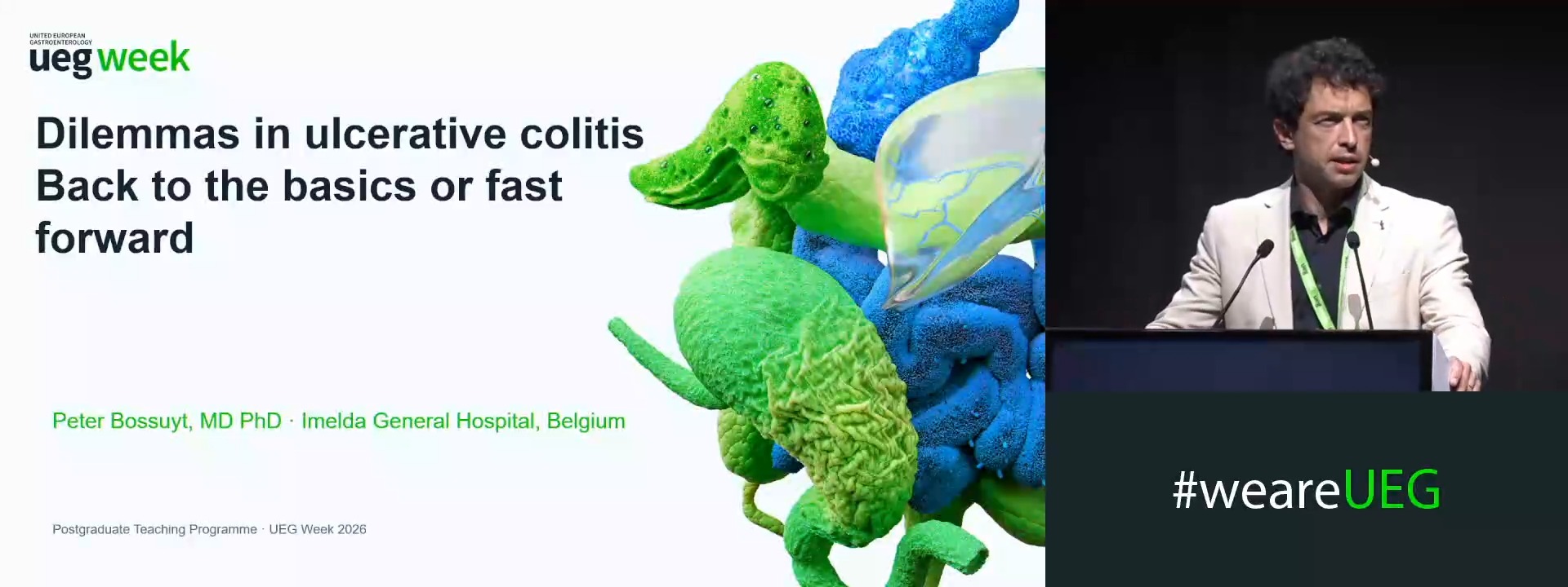
Introduction: Dilemmas in ulcerative colitis: Back to the basics or fast forward
Affiliations
1 Imelda General Hospital, Bonheiden, Belgium
Log in to continue.
This content is part of Gutflix. Log in with your myUEG account, or create one free, to watch it.
Not sure what you can access? Learn more about account types.
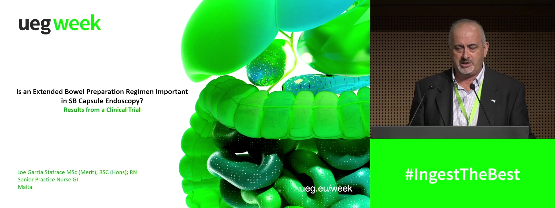
The effectiveness of an extended bowel preparation regimen in capsule endoscopy
Affiliations
1 Mater Dei Hospital, Zabbar, Malta