UEG's content platform for gastroenterology education bringing recordings, courses, guidelines, and more together in one place.
Trending on Gutflix
Rate for better recommendations!
Your feedback helps us show you more of what you like and less of what you dislike!
Log in to continue.
This content is part of Gutflix. Log in with your myUEG account, or create one free, to watch it.
Not sure what you can access? Learn more about account types.
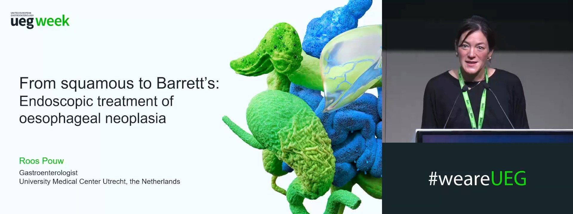
From squamous to Barrett’s: Endoscopic treatment of oesophageal neoplasia
Affiliations
1 Amsterdam UMC, Amsterdam, Netherlands
Log in to continue.
This content is part of Gutflix. Log in with your myUEG account, or create one free, to watch it.
Not sure what you can access? Learn more about account types.
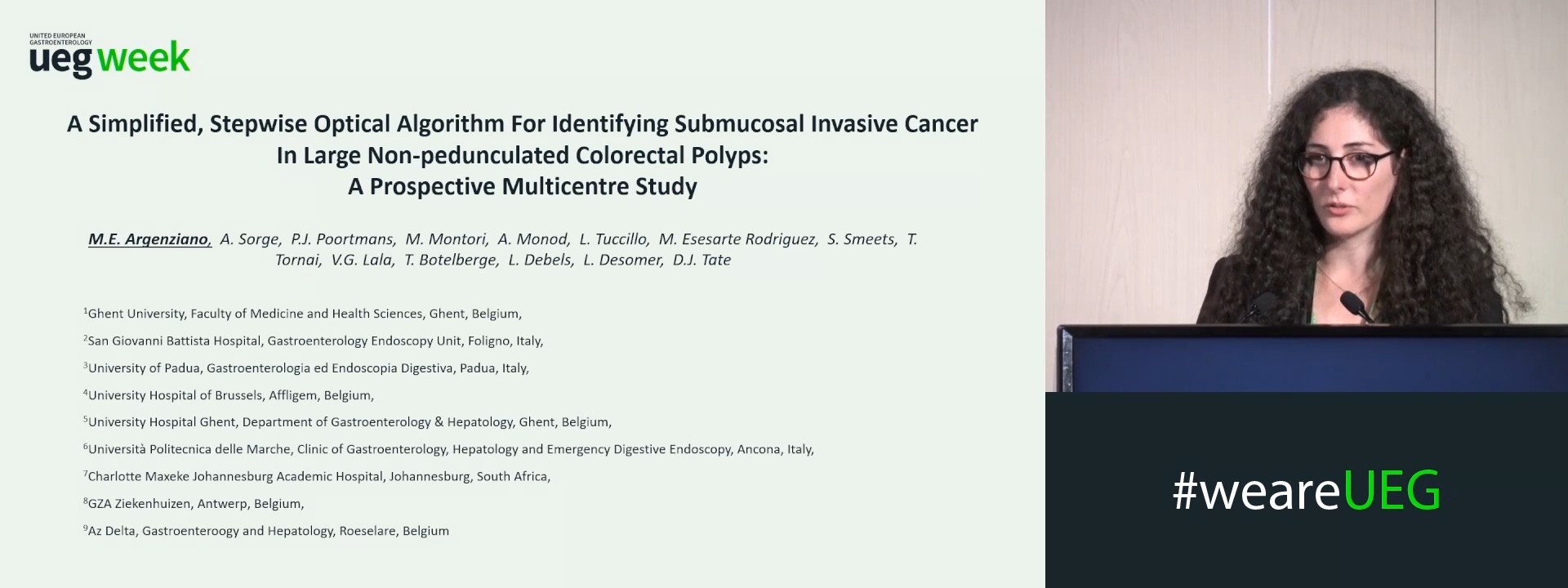
A SIMPLIFIED, STEPWISE OPTICAL ALGORITHM FOR IDENTIFYING SUBMUCOSAL INVASIVE CANCER IN LARGE NON-PEDUNCULATED COLORECTAL POLYPS: A PROSPECTIVE MULTICENTRE STUDY
Maria Eva Argenziano 1,2, Andrea Sorge 3, Pieter Jan Poortmans 1,4,5, Michele Montori 6, Adam Monod 5, Luigi Tuccillo 5, Marisol Esesarte Rodriguez 5, Sander Smeets 5, Tamás Tornai 5, Vikash Goolab Lala 7, Thomas Botelberge 8, Lynn Debels 1,4, Lobke Desomer 1,9, David James Tate 1,5
Affiliations
1 Ghent University, Ghent, Belgium
2 San Giovanni Battista Hospital, Foligno, Italy
3 University of Padua, Padua, Italy
4 University Hospital of Brussels, Affligem, Belgium
5 University Hospital Ghent, Ghent, Belgium
6 Università Politecnica delle Marche, Ancona, Italy
7 Charlotte Maxeke Johannesburg Academic Hospital, Johannesburg, South Africa
8 GZA Ziekenhuizen, Antwerp, Belgium
9 Az Delta, Roeselare, Belgium

Log in to continue.
This content is part of Gutflix. Log in with your myUEG account, or create one free, to watch it.
Not sure what you can access? Learn more about account types.
Gastric cancer
Affiliations
1 Nantes Université, Nantes, France
Log in to continue.
This content is part of Gutflix. Log in with your myUEG account, or create one free, to watch it.
Not sure what you can access? Learn more about account types.
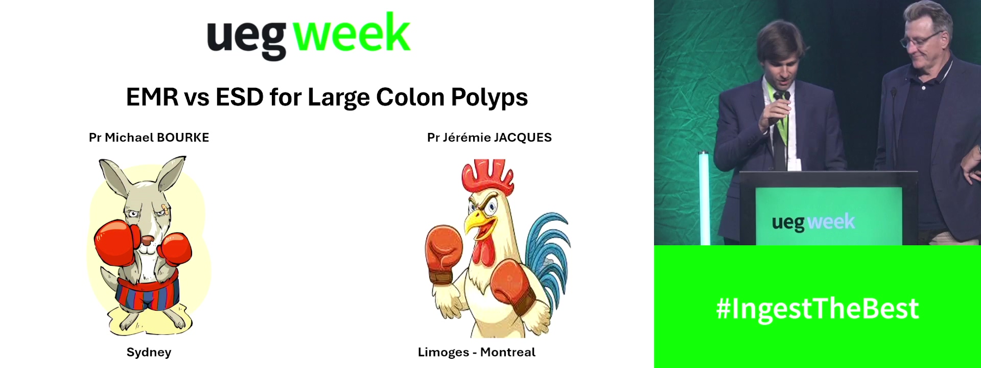
EMR and ESD for large colonic polyps
Affiliations
1 Westmead Hospital Sydney, Westmead, Australia
2 CHU Limoges - Hepato-Gastro-Enterology, CHU Limoges; Limoges/FR, Limoges, France
Log in to continue.
This content is part of Gutflix. Log in with your myUEG account, or create one free, to watch it.
Not sure what you can access? Learn more about account types.
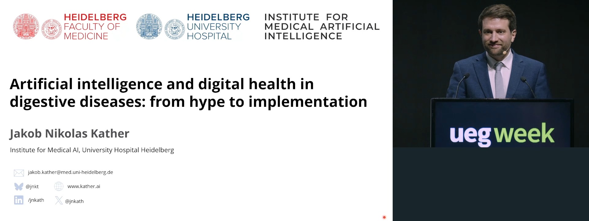
Artificial intelligence and digital health in digestive diseases: From hype to implementation
Affiliations
1 Dresden University of Technology, Dresden, Germany
Log in to continue.
This content is part of Gutflix. Log in with your myUEG account, or create one free, to watch it.
Not sure what you can access? Learn more about account types.
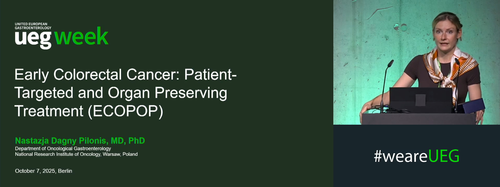
Early colorectal cancer: Patient-targeted and organ preserving treatment (ECOPOP Project)
Affiliations
1 The Maria Sklodowska-Curie Institute - Oncology Institute, Warsaw, Poland