UEG's educational platform for gastroenterology education bringing recordings, courses, guidelines, and more together in one place.
Trending on Gutflix
Rate for better recommendations!
Your feedback helps us show you more of what you like and less of what you dislike!
Log in to continue.
This content is part of Gutflix. Log in with your myUEG account, or create one free, to watch it.
Not sure what you can access? Learn more about account types.
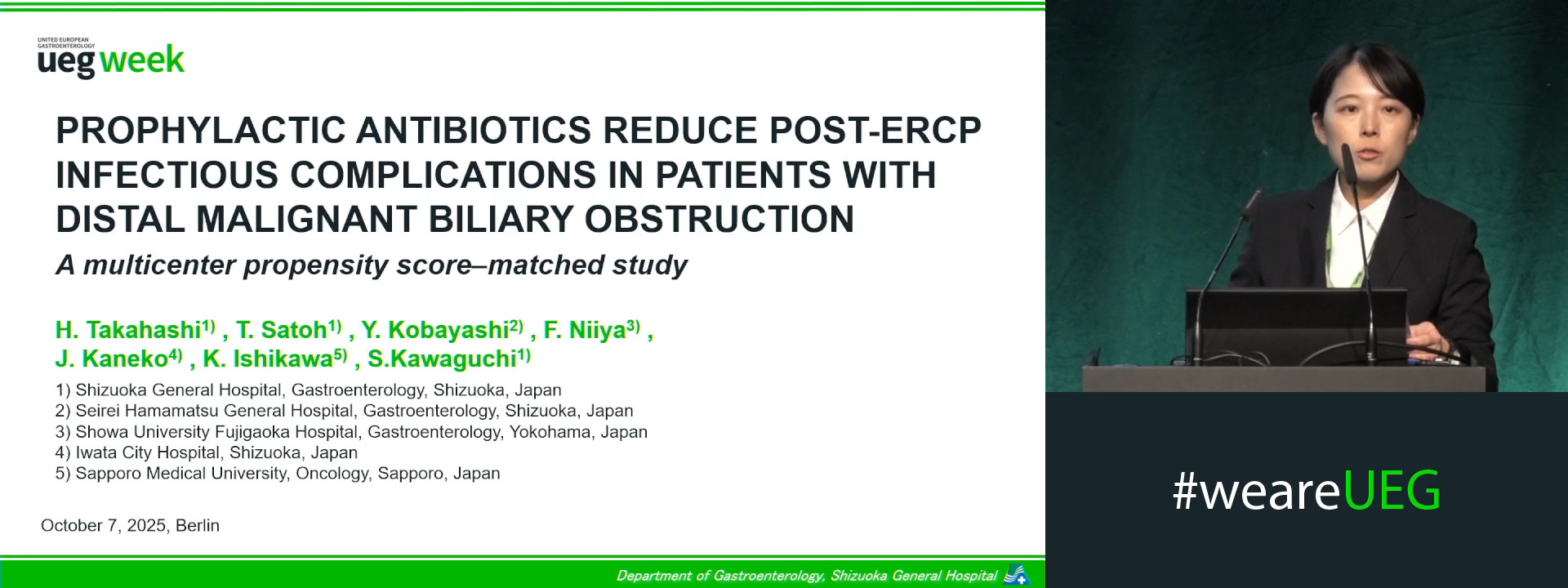
PROPHYLACTIC ANTIBIOTICS REDUCE POST ENDOSCOPIC RETROGRADE CHOLANGIOPANCREATOGRAPHY INFECTIOUS COMPLICATIONS IN PATIENTS WITH DISTAL MALIGNANT BILIARY OBSTRUCTION: A MULTICENTER PROPENSITY SCORE MATCHED STUDY
Haruna Takahashi 1, Tatsunori Satoh 1, Yosuke Kobayashi 2, Fumitaka Niiya 3, Junichi Kaneko 4, Kazuma Ishikawa 5, Shinya Kawaguchi 1
Affiliations
1 Shizuoka General Hospital, Shizuoka, Japan
2 Seirei Hamamatsu General Hospital, Shizuoka, Japan
3 Showa University Fujigaoka Hospital, Yokohama, Japan
4 Iwata City Hospital, Shizuoka, Japan
5 Sapporo Medical University, Sapporo, Japan
Log in to continue.
This content is part of Gutflix. Log in with your myUEG account, or create one free, to watch it.
Not sure what you can access? Learn more about account types.
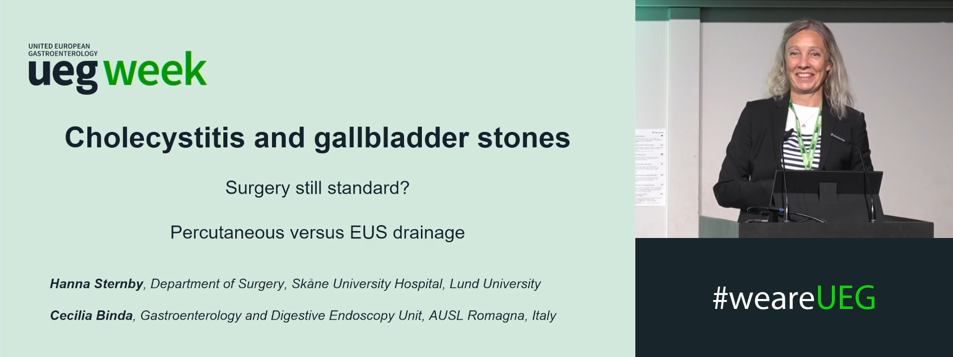
Cholecystitis and gallbladder stones: Is surgery still standard in the era of percutaneous and EUS drainage?
Affiliations
1 AUSL Romagna, Forlì, Italy
2 Institution of Clinical Sciences, Lund, Sweden
Log in to continue.
This content is part of Gutflix. Log in with your myUEG account, or create one free, to watch it.
Not sure what you can access? Learn more about account types.
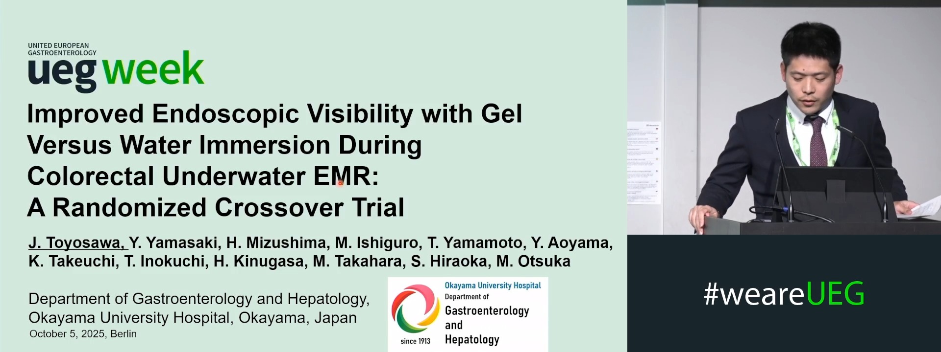
IMPROVED ENDOSCOPIC VISIBILITY WITH GEL VERSUS WATER IMMERSION DURING COLORECTAL UNDERWATER EMR: A RANDOMIZED CROSSOVER TRIAL
Junki Toyosawa 1, Yasushi Yamasaki 1, Hidetaka Mizushima 1, Mikako Ishiguro 1, Takashi Yamamoto 1, Yuki Aoyama 1, Keiko Takeuchi 1, Toshihiro Inokuchi 1, Hideaki Kinugasa 1, Masahiro Takahara 1, Sakiko Hiraoka 1, Motoyuki Otsuka 1
Affiliations
1 Okayama University Hospital, Okayama, Japan
Log in to continue.
This content is part of Gutflix. Log in with your myUEG account, or create one free, to watch it.
Not sure what you can access? Learn more about account types.
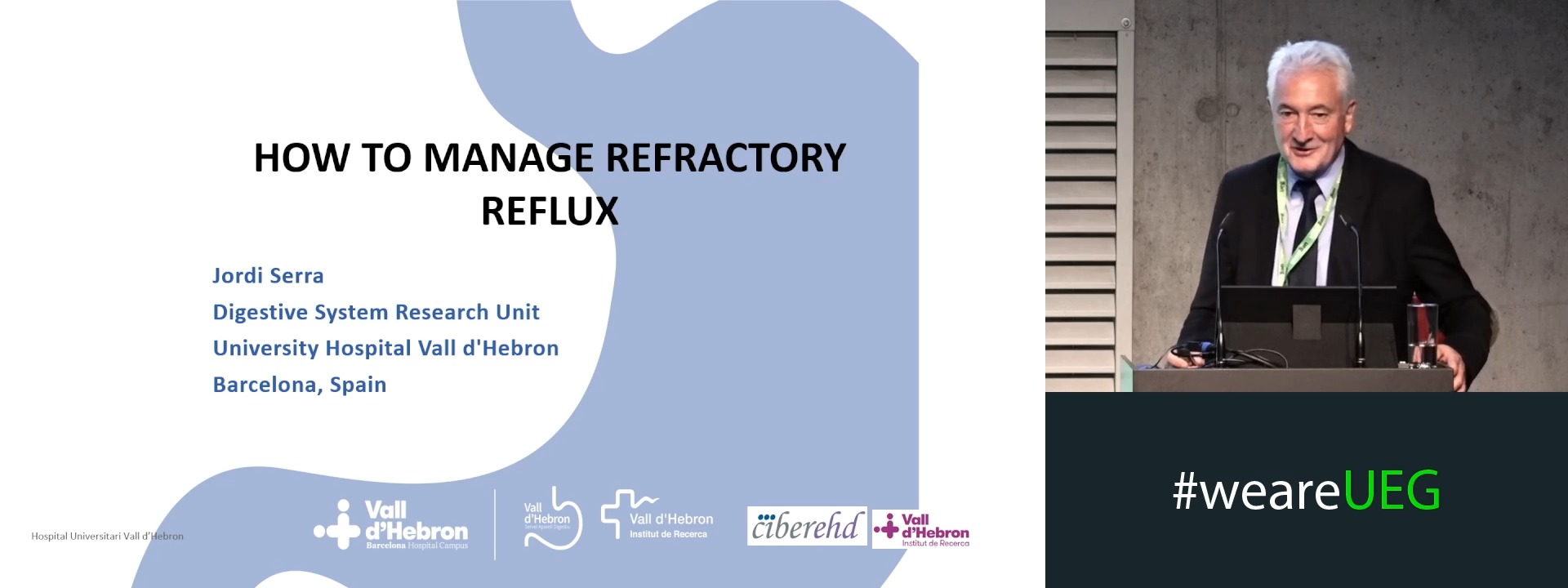
How to manage refractory reflux
Affiliations
1 University Hospital Vall d'Hebron, Barcelona, Spain
Log in to continue.
This content is part of Gutflix. Log in with your myUEG account, or create one free, to watch it.
Not sure what you can access? Learn more about account types.
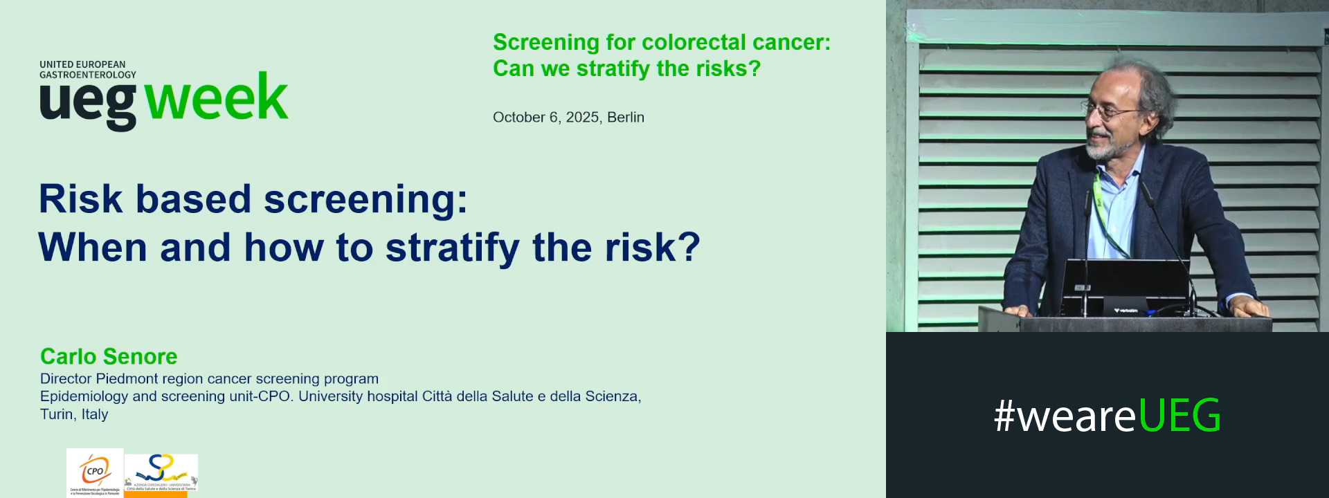
Risk based screening: When and how to stratify the risk?
Affiliations
1 University Hospital Città della Salute e della Scienza, Torino, Italy
Log in to continue.
This content is part of Gutflix. Log in with your myUEG account, or create one free, to watch it.
Not sure what you can access? Learn more about account types.
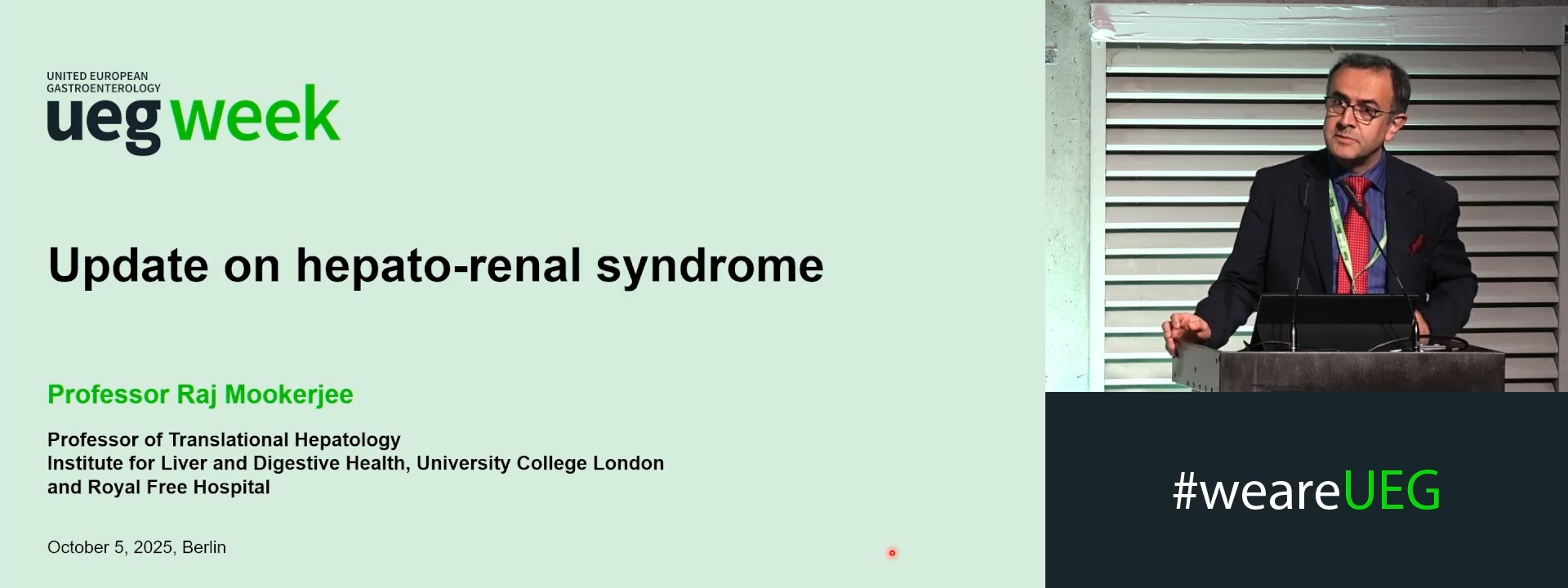
Update on hepatorenal syndrome
Affiliations
1 UCL Institute of Hepatology, University College London, UK, London, United Kingdom
Log in to continue.
This content is part of Gutflix. Log in with your myUEG account, or create one free, to watch it.
Not sure what you can access? Learn more about account types.
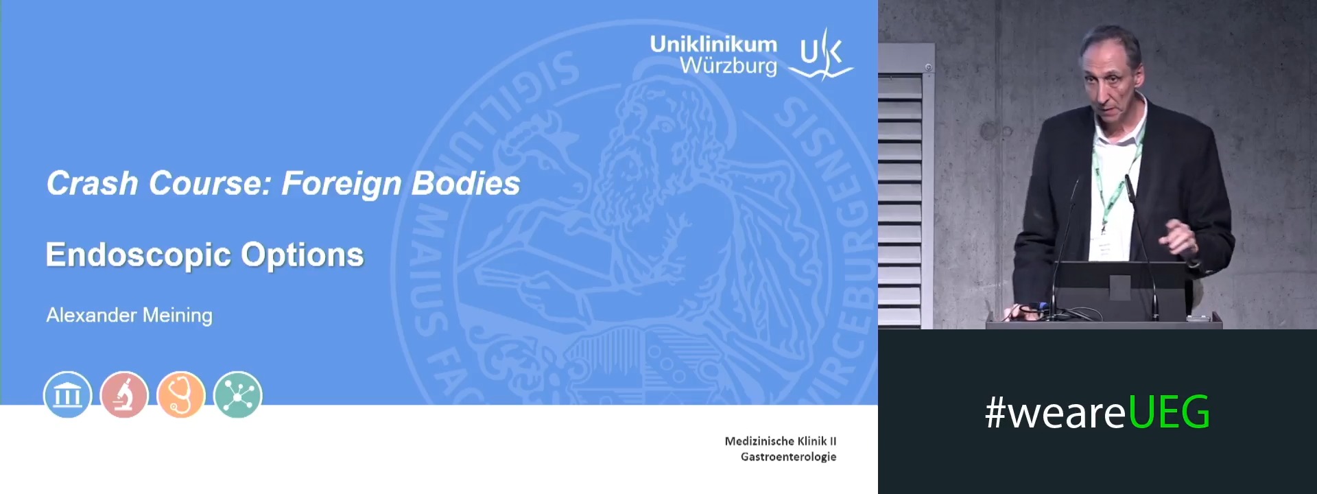
Endoscopic options
Affiliations
1 Bavarian Cancer Research Center (BZKF), Würzburg, Germany