UEG's educational platform for gastroenterology education bringing recordings, courses, guidelines, and more together in one place.
Most popular content
Rate for better recommendations!
Your feedback helps us show you more of what you like and less of what you dislike!
Log in to access this content.
Free for all myUEG account holders. Your access level is set automatically based on your occupation. Medical professionals get full access to all content. If you are a non-medical user, you can only access UEG Week content from congresses you attended.
Not sure what you can access? Learn more about account types.

IMMUNOPROFILING OF T CELL SUBTYPES IN EARLY-ONSET VERSUS LATER-ONSET COLORECTAL TUMORS: INSIGHTS FROM ENDOSCOPICALLY RESECTED SPECIMENS
Naohiko Akimoto 1, Atsushi Tatsuguchi 1, AITOSHI HOSHIMOTO 1, Shun Nakagome 1, Yuka Shimazu 1, Ryosuke Inoue 1, Tsugumi Habu 1, Eriko Koizumi 1, Kazutoshi Higuchi 1, Takayoshi Nishimoto 1, Jun Omori 1, Ryuji Ohashi 1, Masanori Atsukawa 1
1 Nippon Medical School, Graduate School of Medicine, Tokyo, Japan
Conference
Topics
Submission format
Citation
Published
Log in to access this content.
Free for all myUEG account holders. Your access level is set automatically based on your occupation. Medical professionals get full access to all content. If you are a non-medical user, you can only access UEG Week content from congresses you attended.
Not sure what you can access? Learn more about account types.
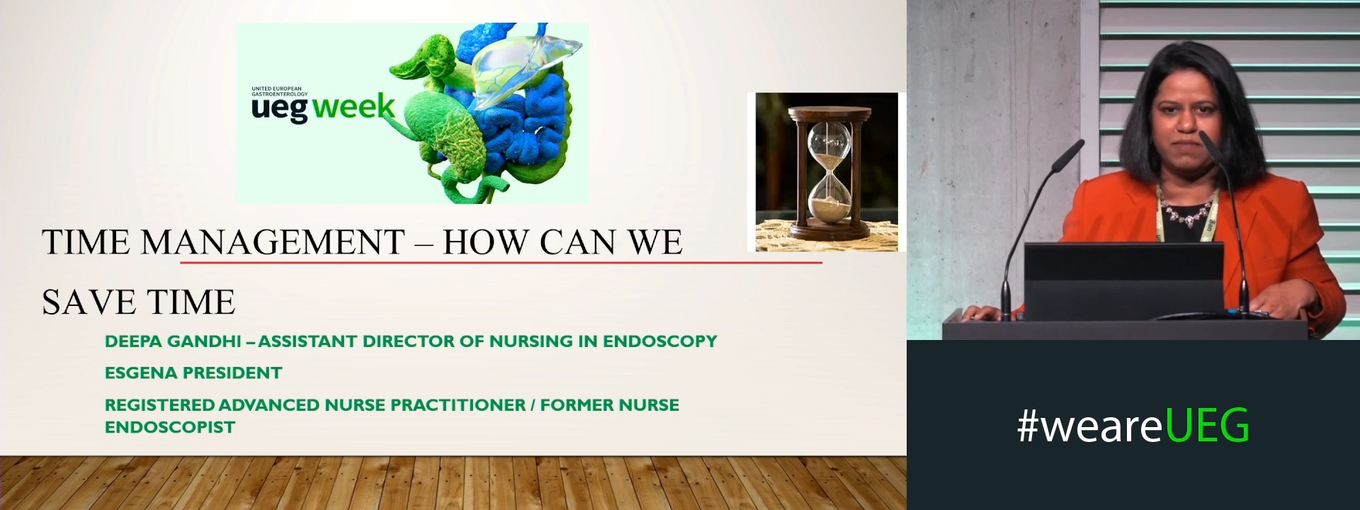
Time management: How can we save time?
1 Mater Misericordiae University Hospital, Dublin, Ireland
Event
Topics
Session
Citation
Published
Log in to access this content.
Free for all myUEG account holders. Your access level is set automatically based on your occupation. Medical professionals get full access to all content. If you are a non-medical user, you can only access UEG Week content from congresses you attended.
Not sure what you can access? Learn more about account types.
Laudatio UEG Lifetime Achievement Awardee
1 IPO Porto, Porto, Portugal
Event
Topics
Session
Citation
Published
Log in to access this content.
Free for all myUEG account holders. Your access level is set automatically based on your occupation. Medical professionals get full access to all content. If you are a non-medical user, you can only access UEG Week content from congresses you attended.
Not sure what you can access? Learn more about account types.
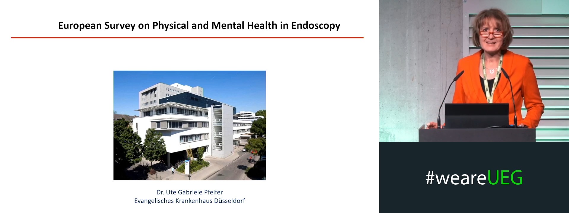
European survey on physical and mental health in endoscopy
1 Evangel. Krankenhaus Düsseldorf Abt. Endoskopie, Düsseldorf, Germany
Event
Topics
Session
Citation
Published
Log in to access this content.
Free for all myUEG account holders. Your access level is set automatically based on your occupation. Medical professionals get full access to all content. If you are a non-medical user, you can only access UEG Week content from congresses you attended.
Not sure what you can access? Learn more about account types.
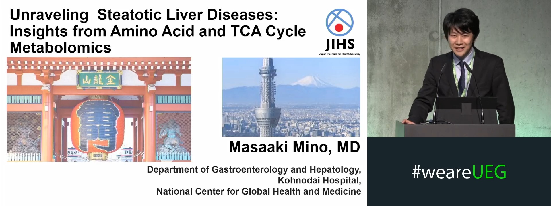
Unraveling Steatotic Liver Diseases: Insights from Amino Acid and TCA Cycle Metabolomics
1 Kohnodai Hospital, National Center for Global Health and Medicine, Kōnodai, Japan
Event
Topics
Citation
Published
Log in to access this content.
Free for all myUEG account holders. Your access level is set automatically based on your occupation. Medical professionals get full access to all content. If you are a non-medical user, you can only access UEG Week content from congresses you attended.
Not sure what you can access? Learn more about account types.
STORE-INDEPENDENT ORAI1 SIGNALING DEFINES BASAL CFTR FUNCTION IN SECRETORY EPITHELIA
Aletta Kata Kiss 1, Arpad Varga 1, Marietta Görög 2, Tamara Madacsy 1, Woo Young Chung 3, Petra Pallagi 1, Viktória Szabó 1, Petra Susánszki 2, Enikő Kúthy-Sutus 1, Szintia Barnai 2, Dániel Varga 4, Péter Bíró 4, Ingrid Hegnes Sendstad 1, Tim Crul 1, Boldizsár Jójárt 1, Bálint Tél 5, Anita Balazs 6, György Lázár 4, Miklós Erdélyi 4, Shmuel Muallem 3, József Maléth 1
1 University of Szeged, Szeged, Hungary|||University of Szeged, Szeged, Hungary|||University of Szeged, Szeged, Hungary
2 University of Szeged, Szeged, Hungary|||University of Szeged, Szeged, Hungary
3 National Institutes of Health, Bethesda, United States
4 University of Szeged, Szeged, Hungary
5 University of Szeged, Szeged, Hungary|||University of Szeged, Szeged, Hungary|||Semmelweis University, Budapest, Hungary
6 Charité Universitätsmedizin Berlin, Berlin, Germany
Event
Submission format
Citation
Published
Log in to access this content.
Free for all myUEG account holders. Your access level is set automatically based on your occupation. Medical professionals get full access to all content. If you are a non-medical user, you can only access UEG Week content from congresses you attended.
Not sure what you can access? Learn more about account types.
Tiny tummies, big problems: Fighting malabsorption in kids
1 University Childrens Hospital Cologne, Cologne, Germany