UEG's content platform for gastroenterology education bringing recordings, courses, guidelines, and more together in one place.
Trending on Gutflix
Rate for better recommendations!
Your feedback helps us show you more of what you like and less of what you dislike!
Log in to continue.
This content is part of Gutflix. Log in with your myUEG account, or create one free, to watch it.
Not sure what you can access? Learn more about account types.
Panel discussion: Quality in endoscopic practice
Log in to continue.
This content is part of Gutflix. Log in with your myUEG account, or create one free, to watch it.
Not sure what you can access? Learn more about account types.
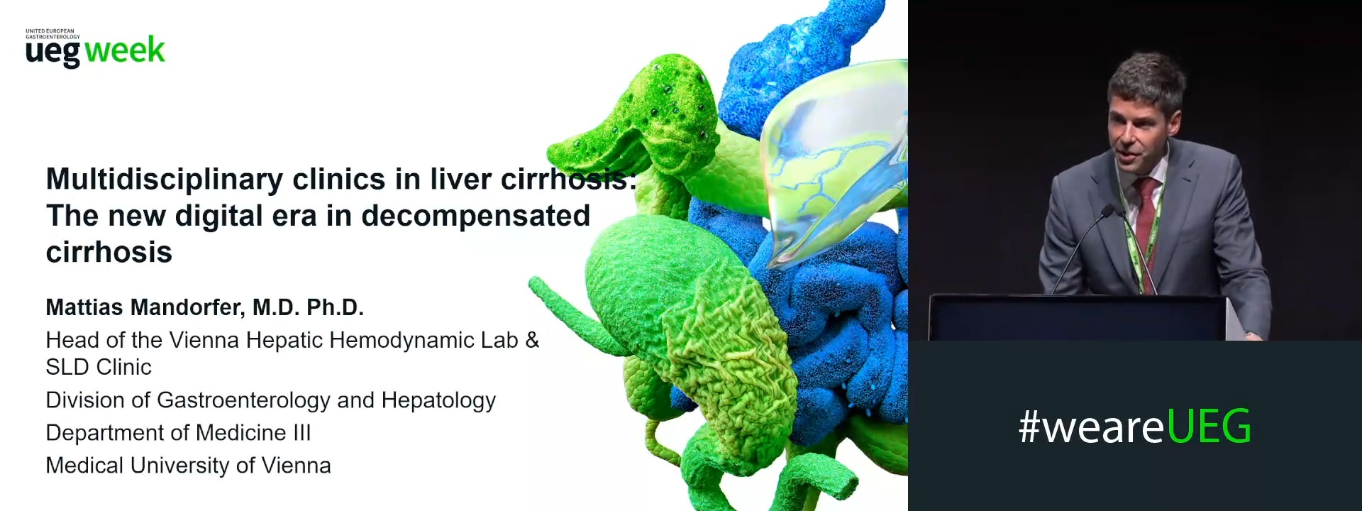
Multidisciplinary clinics in liver cirrhosis: The new digital era in decompensated cirrhosis
Affiliations
1 Medical University of Vienna, Vienna, Austria
Log in to continue.
This content is part of Gutflix. Log in with your myUEG account, or create one free, to watch it.
Not sure what you can access? Learn more about account types.
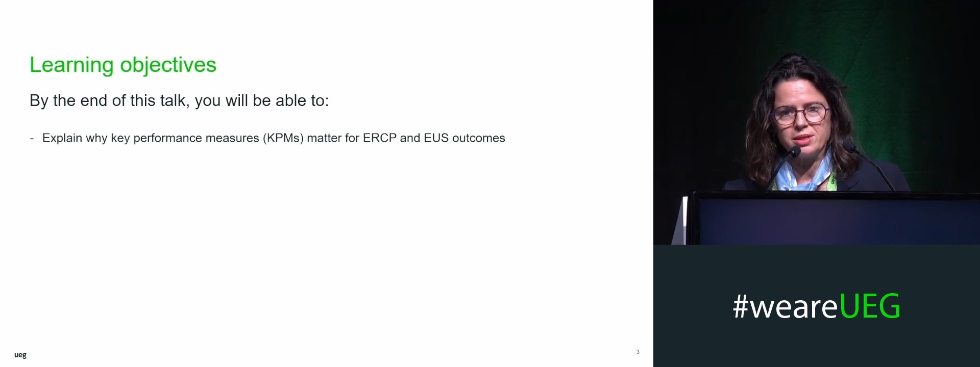
How to disseminate key performance measures in biliopancreatic endoscopy?
Affiliations
1 ERASME University Hospital, Université Libre de Bruxelles, Brussels, Belgium
Log in to continue.
This content is part of Gutflix. Log in with your myUEG account, or create one free, to watch it.
Not sure what you can access? Learn more about account types.
Words of welcome
Affiliations
1 Saarland University Medical Center, Homburg, Germany
2 University of Nottingham, Nottingham, United Kingdom
Log in to continue.
This content is part of Gutflix. Log in with your myUEG account, or create one free, to watch it.
Not sure what you can access? Learn more about account types.
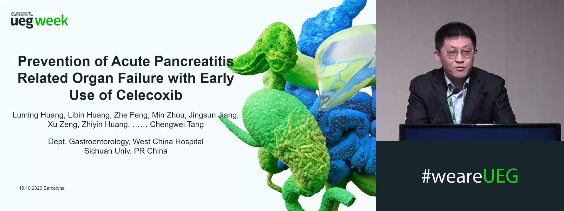
PREVENTION OF ACUTE PANCREATITIS RELATED ORGAN FAILURE WITH EARLY USE OF CELECOXIB -- AN INVESTIGATOR-INITIATED, PROSPECTIVE, MULTI-CENTER, DOUBLE-BLIND, RANDOMIZED, PLACEBO-CONTROLLED CLINICAL TRIAL
Luming Huang 1, Libin Huang 1, Zhe Feng 1, Min Zhou 1, jingsun jiang 1, Xu Zeng 1, Zhiyin Huang 1, Chengwei Tang 1
Affiliations
1 West China Hospital, Sichuan University, Chengdu, China
Log in to continue.
This content is part of Gutflix. Log in with your myUEG account, or create one free, to watch it.
Not sure what you can access? Learn more about account types.
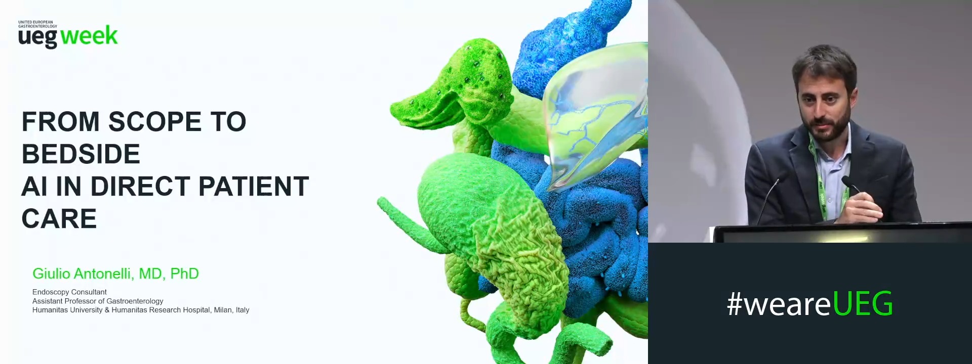
From scope to bedside: AI in direct patient care
Affiliations
1 Ospedale dei Castelli, Rome, Italy