UEG's educational platform for gastroenterology education bringing recordings, courses, guidelines, and more together in one place.
Most popular content
Rate for better recommendations!
Your feedback helps us show you more of what you like and less of what you dislike!
Log in to access this content.
Free for all myUEG account holders. Your access level is set automatically based on your occupation. Medical professionals get full access to all content. If you are a non-medical user, you can only access UEG Week content from congresses you attended.
Not sure what you can access? Learn more about account types.

FECAL CALPROTECTIN AND FECAL ALPHA-1 ANTITRYPSIN – IS THERE A CORRELATION BETWEEN THESE TWO INFLAMMATORY MARKERS?
1 Rabin Medical Center - Beilinson Hospital, Petah Tikva, Israel|||Holon Institute of Technology, Holon, Israel
2 Rabin Medical Center - Beilinson Hospital, Petah Tikva, Israel
Conference
Topics
Submission format
Session
Citation
Published
Log in to access this content.
Free for all myUEG account holders. Your access level is set automatically based on your occupation. Medical professionals get full access to all content. If you are a non-medical user, you can only access UEG Week content from congresses you attended.
Not sure what you can access? Learn more about account types.
Nutrition in coeliac disease for a clinician with Cristian Costas
Topics
Published
Log in to access this content.
Free for all myUEG account holders. Your access level is set automatically based on your occupation. Medical professionals get full access to all content. If you are a non-medical user, you can only access UEG Week content from congresses you attended.
Not sure what you can access? Learn more about account types.
Biliary stones: Diagnostic algorithm and timing
1 ERASME University Hospital, Université Libre de Bruxelles, Brussels, Belgium
Event
Topics
Citation
Published
Log in to access this content.
Free for all myUEG account holders. Your access level is set automatically based on your occupation. Medical professionals get full access to all content. If you are a non-medical user, you can only access UEG Week content from congresses you attended.
Not sure what you can access? Learn more about account types.
Hepatocellular carcinoma
1 HELMHOLTZ ZENTRUM MUENCHEN DEUTSCHES FORSCHUNGSZENTRUM FUER GESUNDHEIT UND UMWELT GMBH, Munich, Germany
Event
Citation
Published
Log in to access this content.
Free for all myUEG account holders. Your access level is set automatically based on your occupation. Medical professionals get full access to all content. If you are a non-medical user, you can only access UEG Week content from congresses you attended.
Not sure what you can access? Learn more about account types.
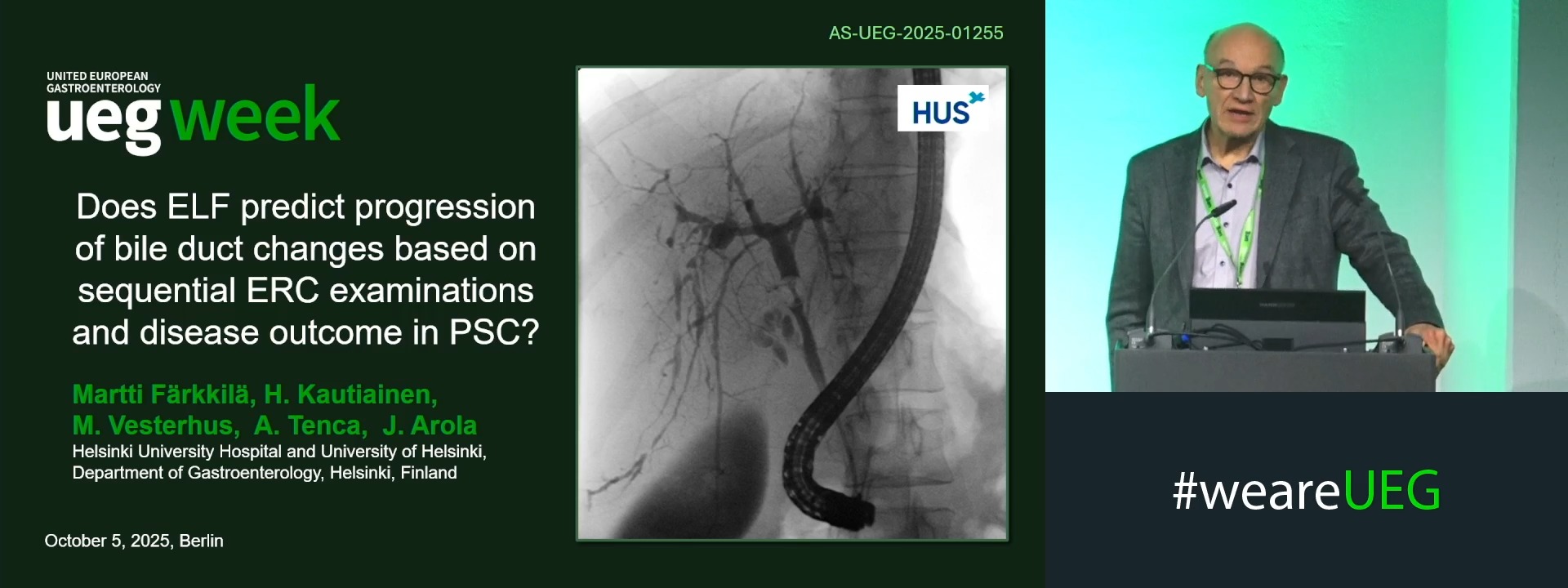
DOES ELF PREDICT PROGRESSION BILE DUCT CHANGES BASED ON SEQUENTIAL ERC EXAMINATIONS AND DISEASE OUTCOME IN PSC?
1 Helsinki University Hospital, Helsinki, Finland
2 University of Eastern Finland, Kuopio, Finland
3 Haraldsplass Deaconess Hospital, Bergen, Norway
4 Clinic of Gastroenterology - Helsinki University Hospital, Helsinki, Finland
5 University of Helsinki, Helsinki, Finland
Event
Submission format
Citation
Published
Log in to access this content.
Free for all myUEG account holders. Your access level is set automatically based on your occupation. Medical professionals get full access to all content. If you are a non-medical user, you can only access UEG Week content from congresses you attended.
Not sure what you can access? Learn more about account types.
ONE-YEAR CHANGES IN ALT AND LSM, BUT NOT IN CAP, PREDICT LONG-TERM LIVER OUTCOMES IN PATIENTS WITH MASLD
Grazia Pennisi 1, Giuseppe Infantino 2, Gabriele Di Maria 2, Emmanuel Tsochatzis 3, Elisabetta Bugianesi 4, Masato Yoneda 5, Ming-Hua Zheng 6, Hannes Hagström 7, Jérôme Boursier 8, Jose Luis Calleja 9, George Boon-Bee Goh 10, Wah-Kheong Chan 11, Rocio Gallego-Durán 12, Arun J Sanyal 13, Victor De Ledinghen 14, Philip Newsome 15, Jian-Gao Fan 16, Laurent Castera 17, Michelle Lai 18, Céline Fournier-Poizat 14, Grace Lai-Hung Wong 5, Angelo Armandi 19, Atsushi Nakajima 20, Wen-Yue Liu 6, Ying Shang 21, Marc de Saint-Loup 22, Elba Llop 23, Kevin Kim Jun Teh 24, Carmen Lara-Romero 25, Amon Asgharpour 26, Sara Mahgoub 15, Mandy Sau-Wai Chan 27, Manuel Romero-Gomez 28, Huapeng Lin 29, Seung Up Kim 30, Terry Cheuk-Fung Yip 5, Adele Tulone 2, Che To Jimmy Lai 5, Boyu Yang 5, Hye-Won Lee 31, Abraham Aregay Desta 32, Giacinta Ciancimino 2, Marco Enea 2, Calogero Camma 33, Vincent Wai Sun Wong 5, Salvatore Petta 34
1 Policlinico Paolo Giaccone, Palermo, Italy
2 University of Palermo, Palermo, Italy
3 UCL Institute of Liver and Digestive Health, London, United Kingdom
4 AOU Città della Salute e della Scienza University of Torino, Torino, Italy, Torino, Italy
5 University of Hong Kong, Hong Kong, China
6 Wenzhou Medical University, Wenzhou, China
7 Department of Upper GI Diseases, Karolinska university hospital Huddinge, Stockholm, Sweden
8 Centre Hospitalier Universitaire, Angers, France
9 University of Madrid, Madrid, Spain
10 University of Singapore, Singapore, Malaysia
11 University of Malaya, Kuala Lumpur, Malaysia
12 University of Seville, Seville, Spain
13 Virginia Commonwealth University School of Medicine, Richmond, United States
14 Echosense, Paris, France
15 University of Birmingham, Birmingham, United Kingdom
16 Shanghai Jiaotong University School of Medicine, Shanghai, China
17 Hôpital Beaujon - Service d’Hépatologie, Hôpital Beaujon; Clichy/FR, Clichy, France
18 Harvard Medical School, Boston, United States
19 University of Turin, Torino, Italy
20 Yokohama City University Hosp. Dept. of Gastroenterology, Yokohama, Japan
21 Karolinska Institutet, Stockholm, Sweden
22 Angers University, Angers, France
23 Gastroenterology, Hospital Puerta de Hierro, Madrid, Spain, Madrid, Spain
24 Changi General Hospital, Singhealth, Singapore, Singapore
25 Punta Europa Hospital, Algeciras, Spain
26 VCU School of Medicine, Richmond, United States
27 Echosens, Paris, France
28 Valme University Hospital University of Seville, Sevilla, Spain
29 University of Shanghai, Shanghai, China
30 Yonsei University College of Medicine, Seoul, Korea, Republic of
31 Yonsei University College Dept. of Internal Medicine, Seoul, Korea, Republic of
32 University of Florence, Florence, Italy
33 UOC Gastroenterologia Ed Epatologia, Palermo, Italy
34 University of Palermo Italy 48.37, Palermo, Italy
Event
Submission format
Citation
Published
Log in to access this content.
Free for all myUEG account holders. Your access level is set automatically based on your occupation. Medical professionals get full access to all content. If you are a non-medical user, you can only access UEG Week content from congresses you attended.
Not sure what you can access? Learn more about account types.
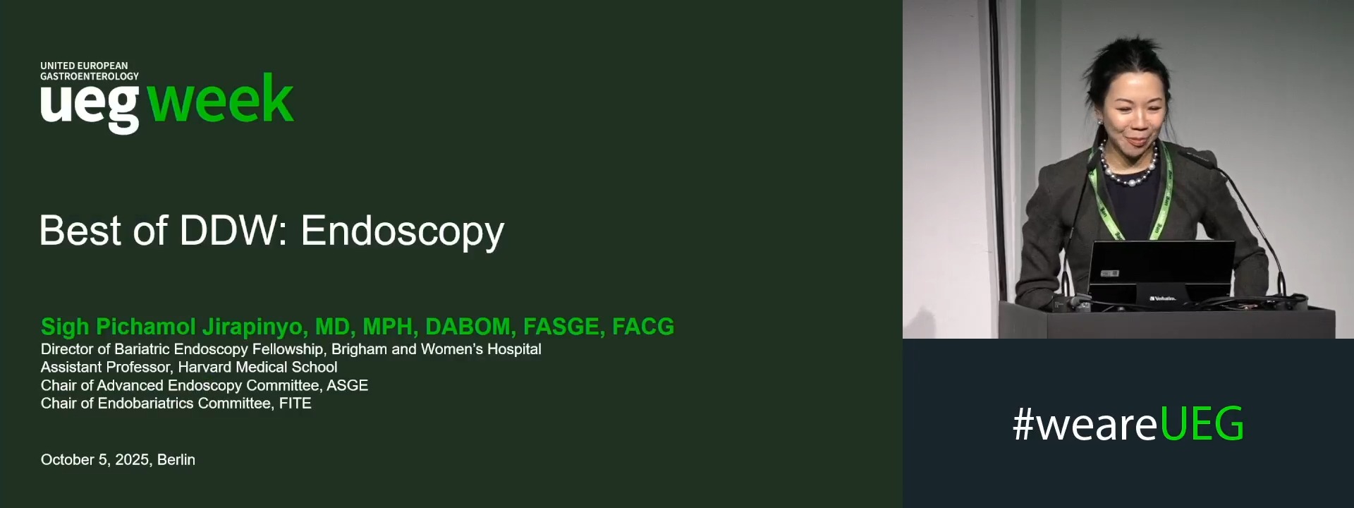
Endoscopy
1 Brigham and Women's Hospital, Boston, United States of America
Event
Session