UEG's content platform for gastroenterology education bringing recordings, courses, guidelines, and more together in one place.
Trending on Gutflix
Rate for better recommendations!
Your feedback helps us show you more of what you like and less of what you dislike!
Log in to continue.
This content is part of Gutflix. Log in with your myUEG account, or create one free, to watch it.
Not sure what you can access? Learn more about account types.
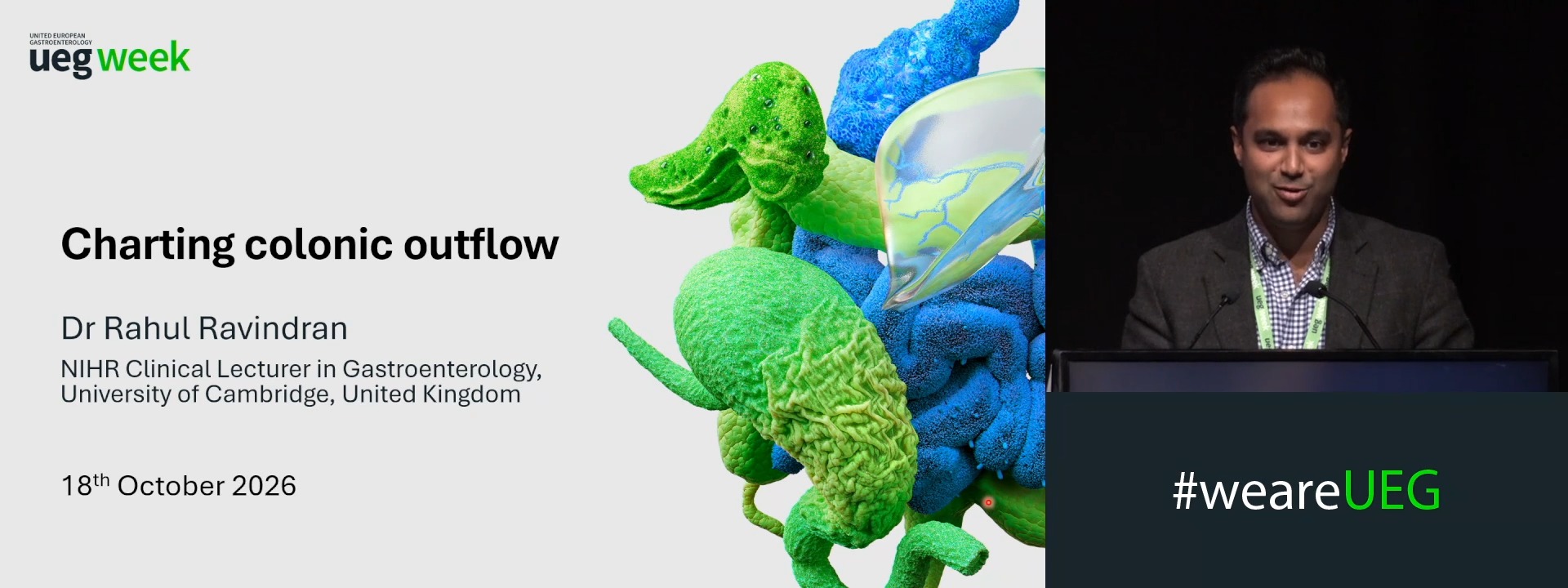
TISSUE FOLDS ORGANISE COLON LYMPHATICS TO CHANNEL INTERSTITIAL OUTFLOW AND PROMOTE RESILIENCE AGAINST INFLAMMATORY SWELLING
Rahul Ravindran 1, Drishya Iyer 2, Daniel Lee 2, Meghan Blitchington 3, Christopher Huckstep 2, Riley Telkamp 3, Kevin Telfer 2, Lillian O'Neal 3, Rachel Mintz 2, Deanna Davis 2, Kibeom Park 2, Nadav Yayon 1, Shashi Kumar 2, Alexandre Gallerand 2, Ahmed Hedayat 2, Changqing Ma 2, Mark Hoofnagle 2, Raphael Blain 4, Adrien Hallou 4, Young-Kwon Hong 5, Simon Travis 4, Matthias Friedrich 4, Huaibin Ko 6, Ryan C. Ungaro 6, Chao Zhou 2, Jean-Frédéric Colombel 6, Fiona Powrie 4, Mark Coles 4, Christopher Buckley 4, Bernd Zinselmeyer 2, Gwendalyn Randolph 2, Rafael Czepielewski 3
Affiliations
1 University of Cambridge, Cambridge, United Kingdom
2 Washington University in St. Louis, Saint Louis, United States
3 Augusta University, Augusta, United States
4 University of Oxford, Oxford, United Kingdom
5 Harvard Medical School, Boston, United States
6 Icahn School of Medicine at Mount Sinai, New York City, United States
Log in to continue.
This content is part of Gutflix. Log in with your myUEG account, or create one free, to watch it.
Not sure what you can access? Learn more about account types.
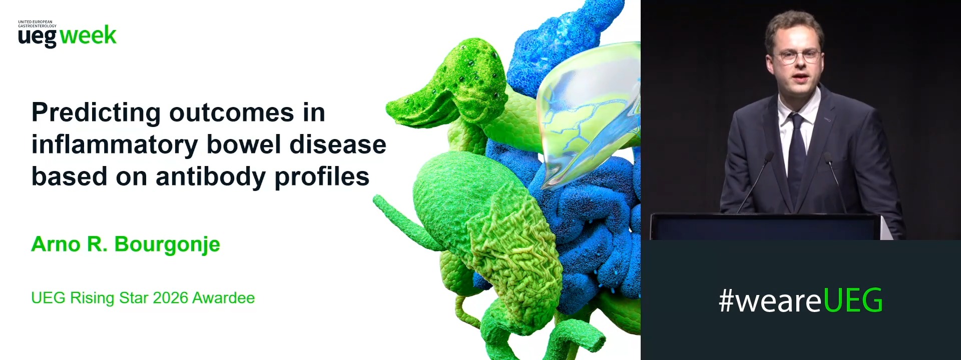
Predicting outcomes based on B-cell profiles
Affiliations
1 University Medical Center of Groningen, Groningen, Netherlands
Log in to continue.
This content is part of Gutflix. Log in with your myUEG account, or create one free, to watch it.
Not sure what you can access? Learn more about account types.
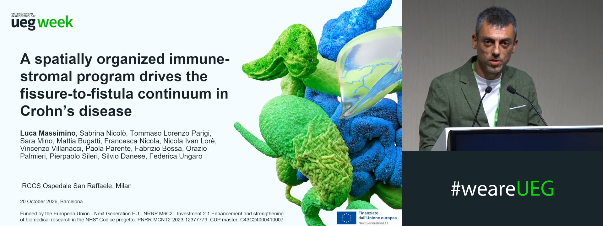
A SPATIALLY ORGANIZED IMMUNE-STROMAL PROGRAM DRIVES THE FISSURE-TO-FISTULA CONTINUUM IN CROHN’S DISEASE
Luca Massimino 1, Sabrina Nicolo' 1, Tommaso Lorenzo Parigi 1, Sara Mino 2, Mattia Bugatti 2, Francesca Nicola 1, Nicola Ivan Lorè 1, Vincenzo Villanacci 2, Paola Parente 3, Fabrizio Bossa 4, Orazio Palmieri 5, Pierpaolo Sileri 1, Silvio Danese 6, Federica Ungaro 1
Affiliations
1 Ospedale San Raffaele, Milano, Italy
2 Institute of Pathology, ASST Spedali Civili and University of Brescia, Brescia, Italy
3 Pathology Unit, Fondazione IRCCS Casa Sollievo della Sofferenza, San Giovanni Rotondo, Italy
4 Fondazione Casa Sollievo della Sofferenza, San Giovanni Rotondo, Italy
5 Division of Gastroenterology, Fondazione IRCCS Casa Sollievo della Sofferenza, San Giovanni Rotondo, Italy
6 Vita-Salute San Raffaele University - IRCCS San Raffaele Scientific Institute, Milan, Italy
Log in to continue.
This content is part of Gutflix. Log in with your myUEG account, or create one free, to watch it.
Not sure what you can access? Learn more about account types.
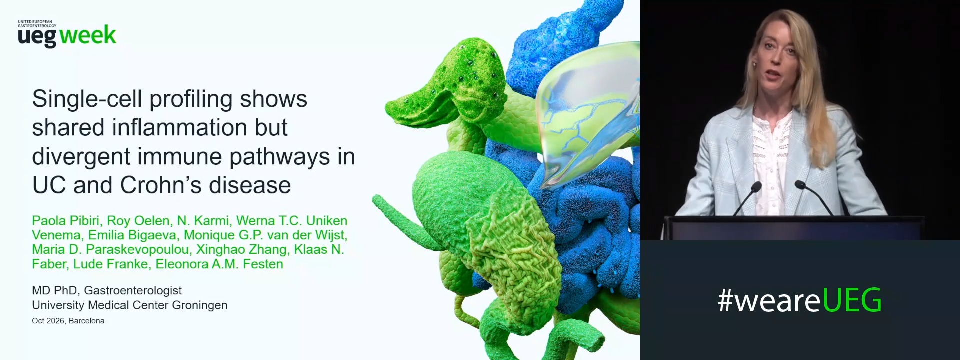
BIOPSY-BASED SINGLE-CELL PROFILING OF ULCERATIVE COLITIS AND CROHN’S DISEASE REVEALS SHARED INFLAMMATORY ARCHITECTURE AND DISEASE-SPECIFIC IMMUNE PATHWAYS
E.A.M. (Noortje) Festen 1, Paola Pibiri 2, Roy Oelen 2, Werna T. C. Uniken Venema 3, Emilia Bigaeva 1, Monique G.P. Van der Wijst 2, Maria Paraskevopoulou 4, Klaas Nico Faber 2
Affiliations
1 UMCG, Groningen, Netherlands
2 University Medical Center Groningen, Groningen, Netherlands
3 Zuyderland Medisch Centrum, Leuven, Belgium
4 Takeda Pharmaceuticals, Acton, United States
Log in to continue.
This content is part of Gutflix. Log in with your myUEG account, or create one free, to watch it.
Not sure what you can access? Learn more about account types.
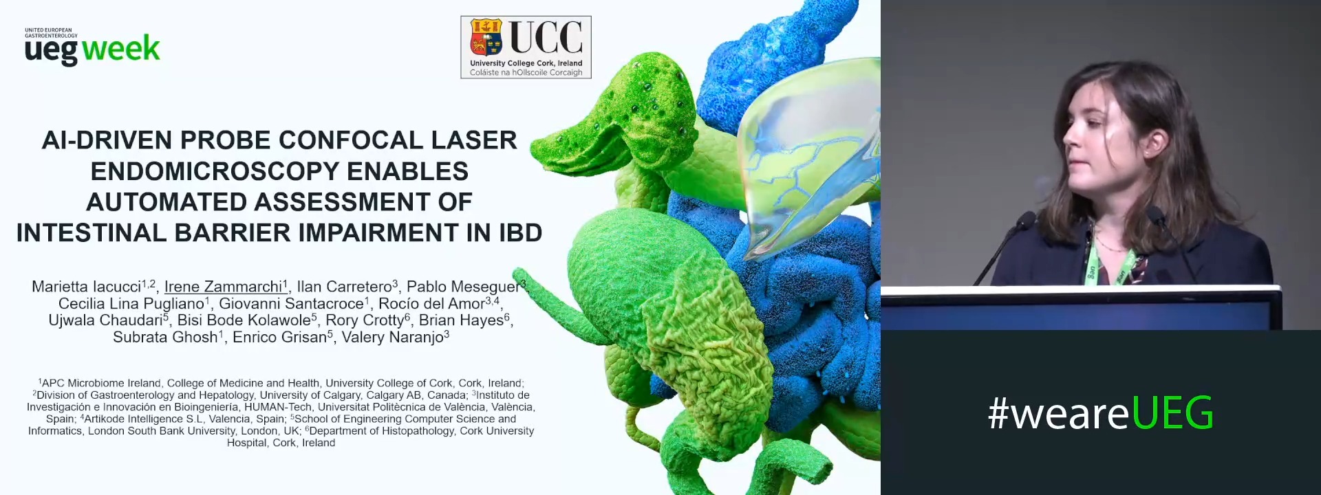
AI-DRIVEN PROBE CONFOCAL LASER ENDOMICROSCOPY ENABLES AUTOMATED ASSESSMENT OF INTESTINAL BARRIER IMPAIRMENT IN IBD
Marietta Iacucci 1,2, Irene Zammarchi 1, Ilan Carretero 3, Pablo Meseguer 3, Cecilia Lina Pugliano 1, Giovanni Santacroce 1, Rocio Del Amor 3,4, Ujwala Chaudhari 5, Bisi Bode Kolawole 5, Rory Crotty 6, Brian Hayes 6, Subrata Ghosh 1, Enrico Grisan 5, Valery Naranjo 3,4
Affiliations
1 University College Cork, Cork, Ireland
2 University of Calgary, Calgary, Canada
3 Universitat Politècnica de València, Valencia, Spain
4 Artikode Intelligence S.L., Valencia, Spain
5 London South Bank University, London, United Kingdom
6 Cork University Hospital, Cork, Ireland