UEG's educational platform for gastroenterology education bringing recordings, courses, guidelines, and more together in one place.
Most popular content
Rate for better recommendations!
Your feedback helps us show you more of what you like and less of what you dislike!
Log in to access this content.
Free for all myUEG account holders. Your access level is set automatically based on your occupation. Medical professionals get full access to all content. If you are a non-medical user, you can only access UEG Week content from congresses you attended.
Not sure what you can access? Learn more about account types.

EVALUATION OF GENES EXPRESSION OF BOTH IMMUNOGLOBULIN SUPERFAMILY DOMAINS AND HIGH MOBILITY GROUP IN DIAGNOSIS AND PROGNOSIS OF COLORECTAL CANCER
Mahmoud El-Bendary 1, Amal Alaa 2, Mustafa Neamatallah 1, Waleed Elnahas 2, Ahmed Mosaad 3, Hatem Elalfy 2, Noha Mahmoud 2, Mohamed Elegezy Moustafa 2
1 Mansoura Faculty of Medicine- Mansoura University, Mansoura, Egypt|||Molecular Genetic Unit of Endemic Hepatogastroenterology and Infectious Diseases (MGUHID), Mansoura, Egypt
2 Mansoura Faculty of Medicine- Mansoura University, Mansoura, Egypt
3 Mansoura Faculty of Medicine-Mansoura University, Mansoura, Egypt
Conference
Topics
Submission format
Session
Citation
Published
Log in to access this content.
Free for all myUEG account holders. Your access level is set automatically based on your occupation. Medical professionals get full access to all content. If you are a non-medical user, you can only access UEG Week content from congresses you attended.
Not sure what you can access? Learn more about account types.
TO BIOPSY, RESECT, OR REFER? RETHINKING THE PRE-REFERRAL PATHWAY AND DECISION-MAKING FOR LARGE NON-PEDUNCULATED COLORECTAL POLYPS
Olaolu Olabintan 1, Homira Ayubi 2, Ali Eqbal 3, Ciaran Judge 4, Jemma Bhoday 4, Marie-Anne Noreillie 5, Shraddha Gulati 2, Noriko SUZUKI 1, Bu Hayee 1, Amyn Haji 4, Andrew Emmanuel 4
1 King’s College Hospital NHS Foundation Trust, London, United Kingdom
2 Kings College Hospital, London, United Kingdom
3 Peninsula Health, Melbourne, Australia
4 King's College Hospital, London, United Kingdom
5 AZ Delta, Roeselare, Belgium
Conference
Topics
Submission format
Citation
Published
Log in to access this content.
Free for all myUEG account holders. Your access level is set automatically based on your occupation. Medical professionals get full access to all content. If you are a non-medical user, you can only access UEG Week content from congresses you attended.
Not sure what you can access? Learn more about account types.
EUS-HGS IS SAFE, EFFECTIVE AND COMPLEMENT WITH ERCP, UNIVERSITY HOSPITAL EXPERIENCE
Mitsuhiro Kida 1, Kai Adachi 1, Taro Hanaoka 1, Akihiko Tamaki 1, Takahiro Kurosu 1, Masafumi Watanabe 2, Toru Kaneko 1, Kosuke Okuwaki 1, Tomohisa Iwai 1, Chikako Kusano 1
1 Kitasato University, Sagamihara, Japan
2 Kitasato University School of Medicine, Sagamihara, Japan
Conference
Topics
Submission format
Session
Citation
Published
Log in to access this content.
Free for all myUEG account holders. Your access level is set automatically based on your occupation. Medical professionals get full access to all content. If you are a non-medical user, you can only access UEG Week content from congresses you attended.
Not sure what you can access? Learn more about account types.
Welcome
1 ESGENA Scientific Secretary, Ulm, Germany
2 Mater Misericordiae University Hospital, Dublin, Ireland
Event
Topics
Session
Citation
Published
Log in to access this content.
Free for all myUEG account holders. Your access level is set automatically based on your occupation. Medical professionals get full access to all content. If you are a non-medical user, you can only access UEG Week content from congresses you attended.
Not sure what you can access? Learn more about account types.
DISTINCT GUT MICROBIAL PROFILES IN LYNCH SYNDROME CARRIERS AND AVERAGE-RISK INDIVIDUALS: IMPLICATIONS FOR COLORECTAL CANCER SURVEILLANCE
Sandra Taboada 1, Marta Malagón 2, Lia Oliver 2, L Jesús Garcia-Gil 3, Mariona Serra-Pagès 2, Xavier Aldeguer Mante 4, Anna Bahí 5, REBECA MOREIRA DE ABREU 6, Antoni Castells 6, Teresa Ocaña 6, Joaquín Castillo 6, Jordi Guardiola 7, Núria Dueñas 8, Virginia Piñol 9, Marta Pineda 8, Gabriel Capellà 8, Sara Ramió-Pujol 2, Laura Baldo 10, Francesc Balaguer 6, Joan Brunet 11
1 GoodGut, Girona, Spain|||University of Girona, Girona, Spain|||University of Barcelona, Barcelona, Spain
2 GoodGut, Girona, Spain
3 University of Girona, Girona, Spain
4 Dr. Josep Trueta University Hospital, Girona, Spain
5 Biomedical Research Institute of Girona, Salt, Spain
6 Hospital Clínic of Barcelona, Barcelona, Spain
7 Hospital Universitario de Bellvitge, L'Hospitalert de LLobregat (Barcelona), Spain
8 Catalan Institute of Oncology, Hospitalet de Llobregat, Spain
9 Dr. Josep Trueta University Hospital, Girona, Spain|||Universitat de Girona, Girona, Spain
10 University of Barcelona, Barcelona, Spain|||Institute for Research on Biodiversity (IRBio), Barcelona, Spain
11 Catalan Institute of Oncology, Girona, Spain|||Catalan Institute of Oncology, Hospitalet de Llobregat, Spain
Conference
Topics
Submission format
Citation
Published
Log in to access this content.
Free for all myUEG account holders. Your access level is set automatically based on your occupation. Medical professionals get full access to all content. If you are a non-medical user, you can only access UEG Week content from congresses you attended.
Not sure what you can access? Learn more about account types.
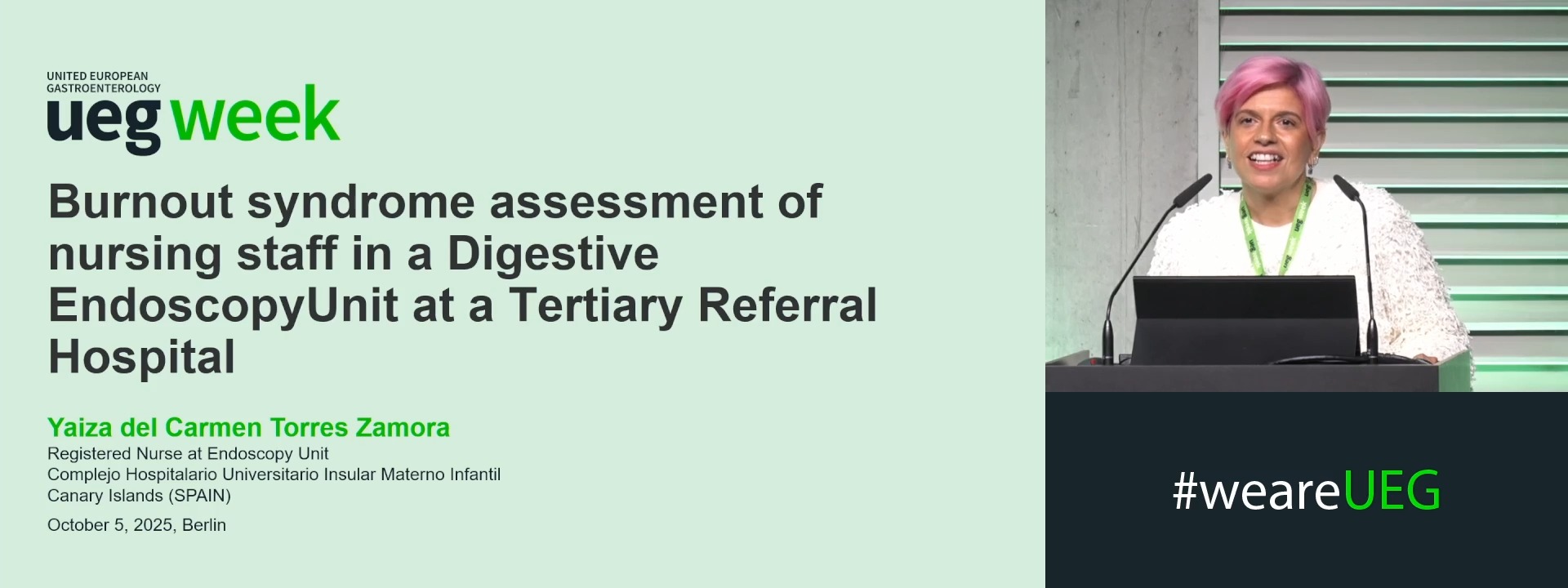
BURNOUT SYNDROME ASSESSMENT OF NURSING STAFF IN A DIGESTIVE ENDOSCOPY UNIT AT A TERTIARY REFERRAL HOSPITAL
1 Complejo Hospitalario Insular Materno Infantil, Las Palmas de Gran Canaria, Spain
Event
Topics
Submission format
Citation
Published
Log in to access this content.
Free for all myUEG account holders. Your access level is set automatically based on your occupation. Medical professionals get full access to all content. If you are a non-medical user, you can only access UEG Week content from congresses you attended.
Not sure what you can access? Learn more about account types.
THE USE OF A NOVEL HEAMOSTATIC PEPTIDE GEL IN THE MANAGEMENT OF ENDOSCOPIC PAPILLECTOMY- RELATED BLEEDING: A CASE SERIES
Stefano Fabbri 1, Cecilia Binda 1, Davide Trama 1, Giacomo Marcozzi 2, Gabriele Rancatore 3, Aurelio Mauro 4, Marco Spadaccini 2, Alessandro Fugazza 2, Chiara Coluccio 1, Paolo Giuffrida 1, Alessandro Repici 2, Ilaria Tarantino 3, Andrea Anderloni 4, Carlo Fabbri 1
1 AUSL Romagna, Forlì, Italy
2 Humanitas Research Hospital, Rozzano, Italy
3 ISMETT, Palermo, Italy
4 Fondazione I.R.C.C.S. Policlinico San Matteo, Pavia, Italy
Conference
Topics
Submission format
Session