UEG's content platform for gastroenterology education bringing recordings, courses, guidelines, and more together in one place.
Trending on Gutflix
Rate for better recommendations!
Your feedback helps us show you more of what you like and less of what you dislike!
Log in to continue.
This content is part of Gutflix. Log in with your myUEG account, or create one free, to watch it.
Not sure what you can access? Learn more about account types.
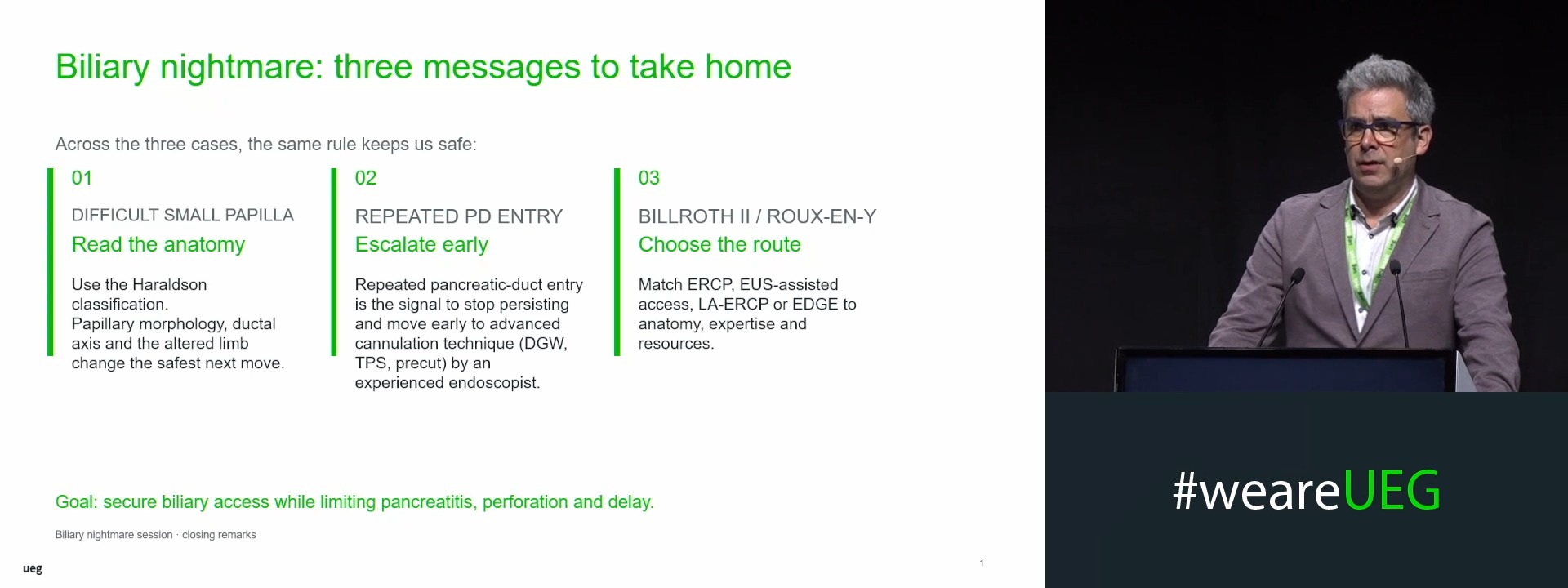
Summary: Are you stuck in the biliary cannulation nightmare: Wake up, here comes the sun
Affiliations
1 Semmelweis University, Budapest, Hungary
Log in to continue.
This content is part of Gutflix. Log in with your myUEG account, or create one free, to watch it.
Not sure what you can access? Learn more about account types.
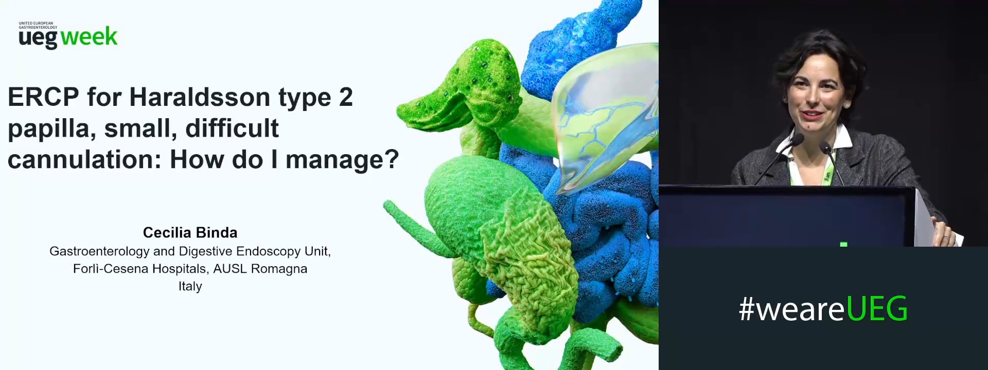
Video case: ERCP for Haraldsson type 2 papilla, small, difficult cannulation: How do I manage?
Affiliations
1 AUSL Romagna, Forlì, Italy
Log in to continue.
This content is part of Gutflix. Log in with your myUEG account, or create one free, to watch it.
Not sure what you can access? Learn more about account types.
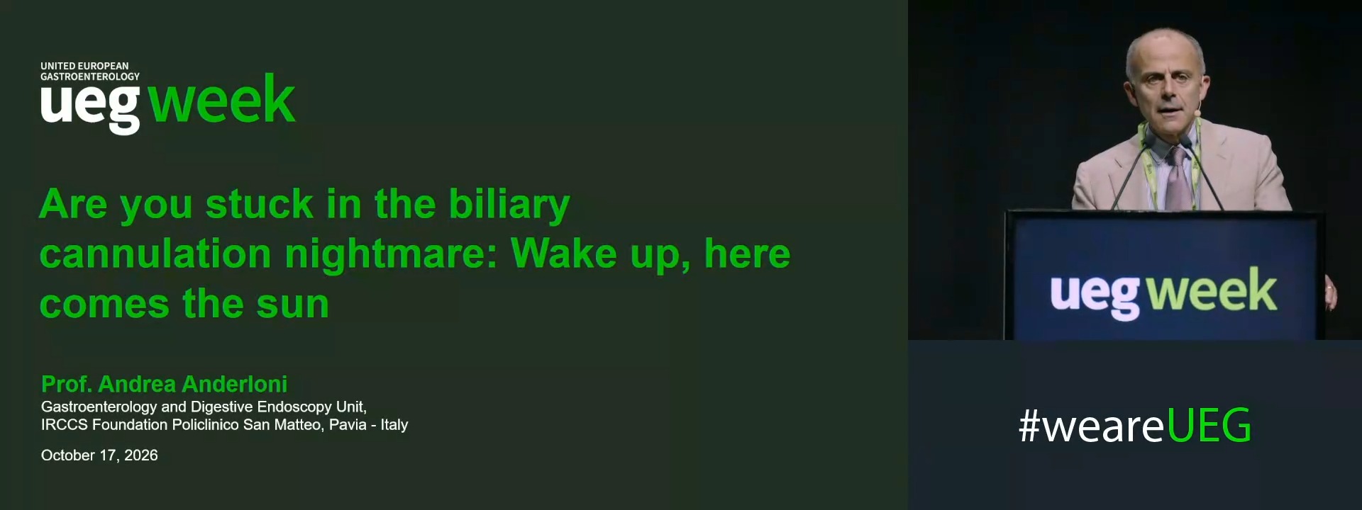
Introduction: Are you stuck in the biliary cannulation nightmare: Wake up, here comes the sun
Affiliations
1 San Matteo Hospital, Pavia, Italy
Log in to continue.
This content is part of Gutflix. Log in with your myUEG account, or create one free, to watch it.
Not sure what you can access? Learn more about account types.
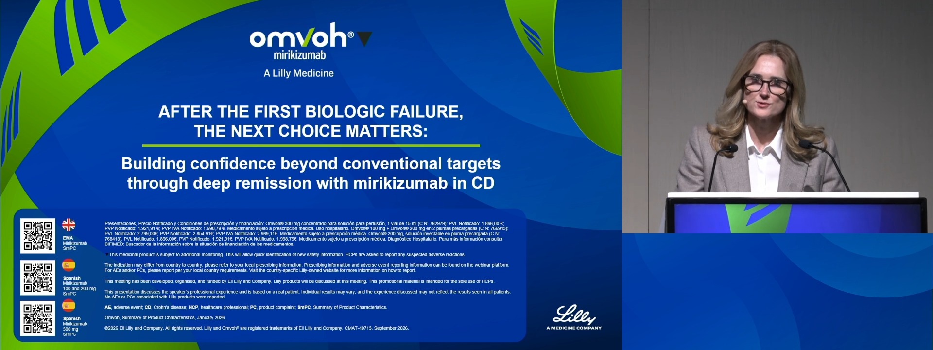
After the first biologic failure, the next choice matters: Building confidence beyond conventional targets through deep remission with mirikizumab in CD (Eli Lilly and Company) (Complete Session)
Affiliations
1 Translational Gastroenterology Unit, Oxford University Hospital, Oxford, United Kingdom
2 University Hospitals Leuven, Leuven, Belgium
3 Department of Internal Medicine I, University Hospital Schleswig-Holstein, Kiel University, Kiel, Germany
Log in to continue.
This content is part of Gutflix. Log in with your myUEG account, or create one free, to watch it.
Not sure what you can access? Learn more about account types.
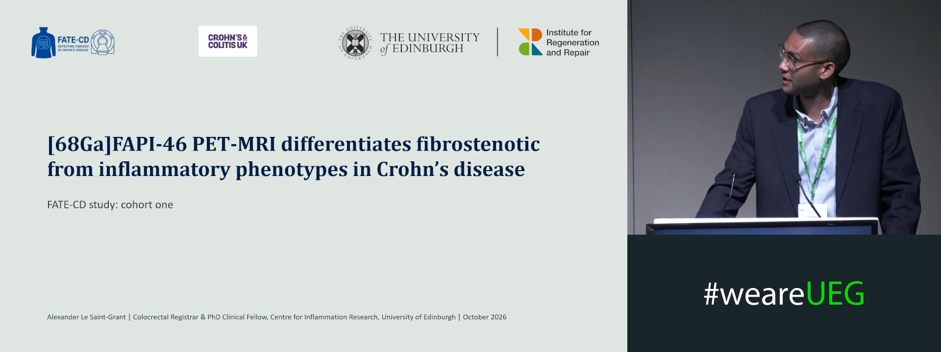
[68GA]FAPI PET-MRI QUANTIFIES FIBROBLAST ACTIVITY BURDEN AND DIFFERENTIATES FIBROSTENOSIS FROM AN INFLAMMATORY PHENOTYPE IN CROHN’S DISEASE
Alexander Le Saint-Grant 1,2, Solomon Xi Xiang Ong 1,2, Calum Worsley 3, Lucy Whitaker 1, Michelle Rooney 2, Nicholas T. Ventham 3, Scott Semple 2, Marc Dweck 2,4, Dilip Patel 3,2, Gwo-Tzer Ho 1,5, David Newby 2,4, Rahul Kalla 2,1
Affiliations
1 Institute for Regeneration and Repair, University of Edinburgh, Edinburgh, United Kingdom
2 Edinburgh Imaging Facility, University of Edinburgh, Edinburgh, United Kingdom
3 NHS Lothian, Edinburgh, United Kingdom
4 University of Edinburgh, Edinburgh, United Kingdom
5 University of Glasgow, Glasgow, United Kingdom
Log in to continue.
This content is part of Gutflix. Log in with your myUEG account, or create one free, to watch it.
Not sure what you can access? Learn more about account types.
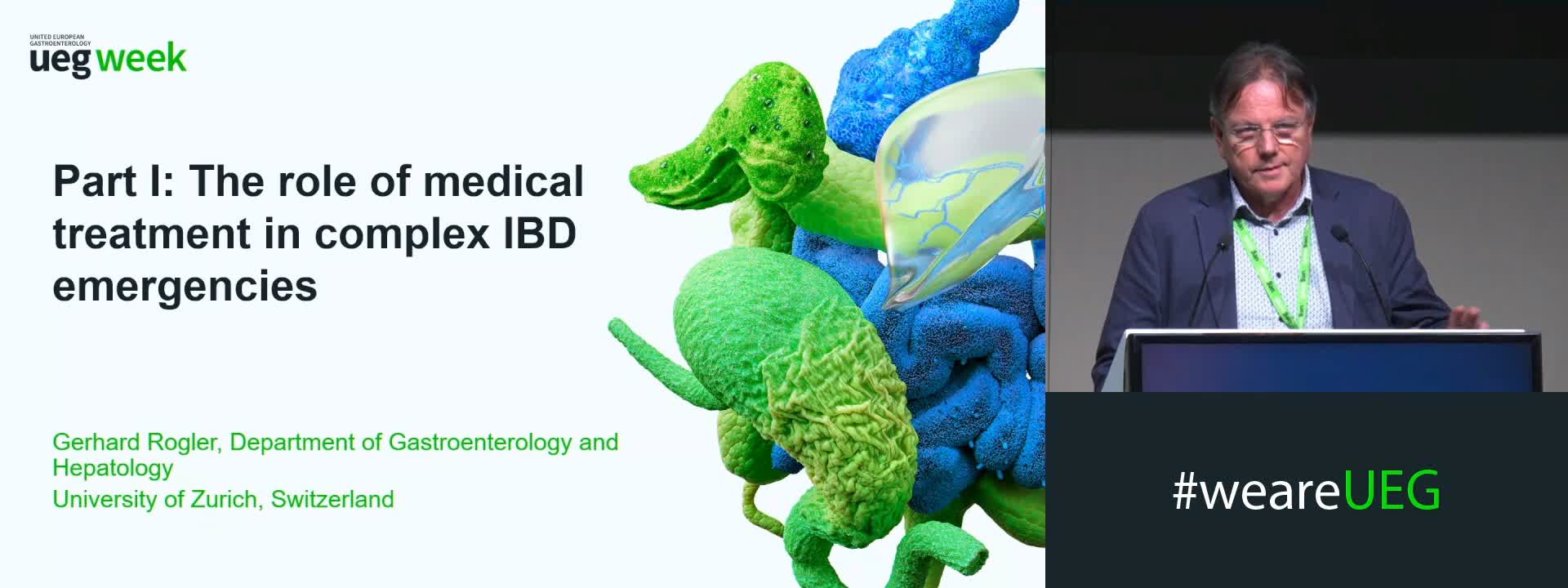
Medical treatment or surgery: What is the solution to complex IBD emergencies?
Affiliations
1 UniversitätsSpital Zürich, Zurich, Switzerland
2 IJsselland Hospital, Capell a/d IJssel, Netherlands
Log in to continue.
This content is part of Gutflix. Log in with your myUEG account, or create one free, to watch it.
Not sure what you can access? Learn more about account types.
Endoscopy: When and how?
Affiliations
1 Katholieke Universiteit Leuven, Leuven, Belgium