UEG's educational platform for gastroenterology education bringing recordings, courses, guidelines, and more together in one place.
Most popular content
Rate for better recommendations!
Your feedback helps us show you more of what you like and less of what you dislike!
Log in to access this content.
Free for all myUEG account holders. Your access level is set automatically based on your occupation. Medical professionals get full access to all content. If you are a non-medical user, you can only access UEG Week content from congresses you attended.
Not sure what you can access? Learn more about account types.
Introduction
In contrast to mucosal T1 rectal cancers with an R0 rate of up to 92% at centers, R0 resection may drop to 30% in case of deep sm infiltration due to thermal artefacts at the basal resection margin. TME often follows as completion surgery. Deep sm infiltration as isolated risk factor excluding L+, V+, G3 and Bd2-3 seems associated with a low risk of LN metastases of only 2.6%. Endoscopic Intermuscular Dissection (EID) is a new flexible endoscopic dissection technique with separation of the circumferential and longitudinal muscular layer. EID ideally provides a specimen incl. the entire submucosa and inner circumferential mp layer. 12 sequential EIDs were compared to 53 historical rectal ESDs with HGIEN or rectal cancer as hp. result.
Aims & Methods
Lesions with biopsy proven ADC or HGIEN of the rectum without endoscopic, EUS or clinical signs of deep local infiltration. 2 pat. included after neo-adjuvant RT/Ch th. 24 and 26 mo before with complete response and refusing definitive surgery. Both with local flat polyps (SSA (1) hyperplastic polyp / LGIEN adenoma (2). Endoscopy, EUS, MRI, CT and rectal digital examination without suspicion of local malignant infiltration. Pts were offered to prospectively undergo EID after extensive informed consent within a prospective protocol. All procedures were carried out under gen. anesthesia by an exp. interv. endoscopist with >1000 ESDs. Results of EID were compared to a historical collective of 53 cases with rectal ESD and a histological result of rectal cancer or HGIEN (08-2018 to 12-2021).
Results
From 11-2023 to 09-2024, 12 EIDs were carried out in 12 pts (6 f/ 6 m; mean 63 y; 34-83y). 09/12 lower rectum, 1/12 middle, 1/12 lower and middle, 1/12 upper rectum. 'En bloc' resection rate was 100%, R0 resection rate 89% for neoplasia / HGIEN. Histopathology: 3x ADC G2, 6x HGIEN, 3x LGIEN including both cases with neodajuvant RT/Ch. R0 resection was achieved in 2 of the 3 ADCs. Both with deep sm infiltration without any other risk factors. Tumor board: close clin. & imaging FU. In the 3rd ADC: TU infiltration of inner muscular layer (T2). Pat. underwent uneventful TME 10 d after EID. Clinical FU in all others. Length of hosp. stay 7.1 days. No serious major complications. All pts. closely followed with uneventful FU.
In the historical ESD group R0 resection was achieved in 100% of the HGIEN cases (45/45) but only in 3/8 lesions (37.5%) of cases with adenocarcinoma.
Conclusion
EID seems a new perspective for the endosc. resection of T1 rectal cancers and suspicious polyps in the rectum. More experience and a comparison to surgical local procedures (TAMIS etc.) is necessary for the final assessment of EID as new promising endoscopic technique.
References

ENDOSCOPIC INTERMUSCULAR DISSECTION (EID) AS NEW PERSPECTIVE FOR T1 RECTAL CANCERS AND HIGH-RISK LESIONS IN THE RECTUM - FIRST COMPARISON OF 12 EID CASES TO 53 LESIONS WITH A HISTOLOGICAL RESULT OF CANCER OR HGIEN RESECTED BY SUBMUCOSAL DISSECTION (ESD)
1 Charité University Medicine Berlin, Berlin, Germany
2 Vivantes Klinikum im Friedrichshain, Berlin, Germany
Conference
Topics
Submission format
Session
Citation
Published
Log in to access this content.
Free for all myUEG account holders. Your access level is set automatically based on your occupation. Medical professionals get full access to all content. If you are a non-medical user, you can only access UEG Week content from congresses you attended.
Not sure what you can access? Learn more about account types.
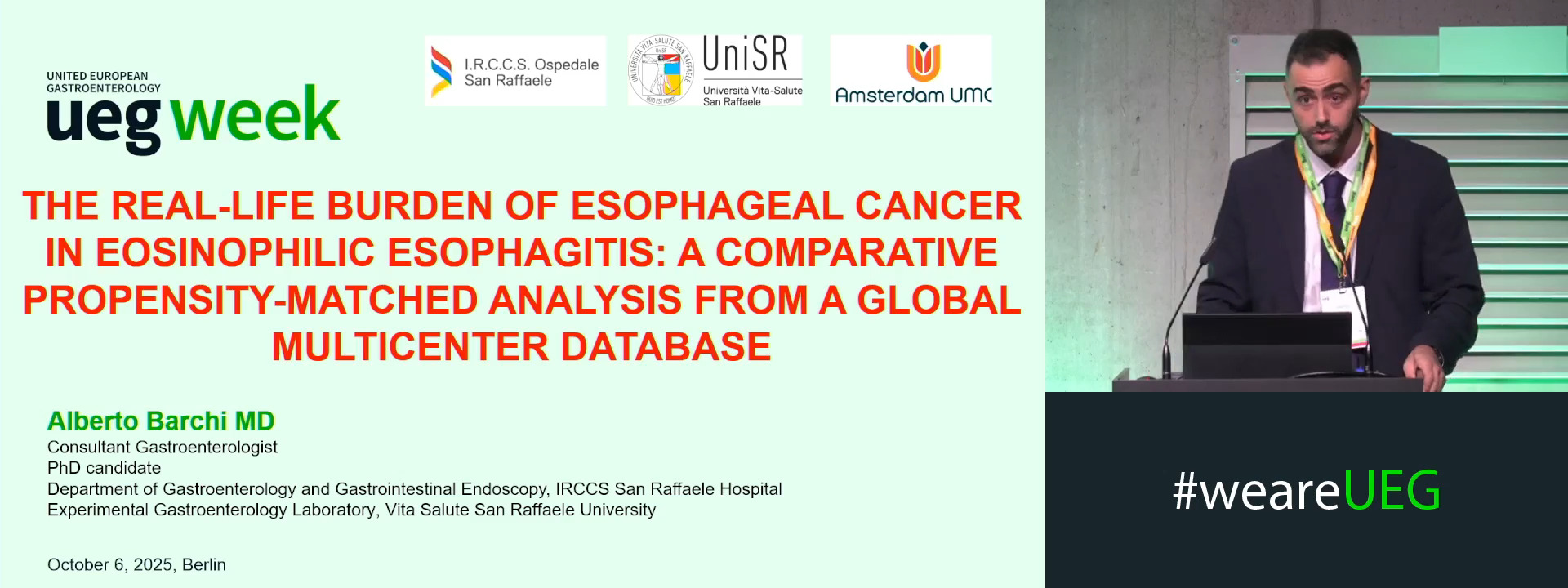
THE REAL-LIFE BURDEN OF ESOPHAGEAL CANCER IN EOSINOPHILIC ESOPHAGITIS: A COMPARATIVE PROPENSITY-MATCHED ANALYSIS FROM A GLOBAL MULTICENTER DATABASE
Alberto Barchi 1, Antonio Ventimiglia 2, Marina Girelli 2, Francesca Rusconi 3, Sandro Passaretti 2, Silvio Danese 2, Pierfrancesco Visaggi 4, Edoardo Vincenzo Savarino 5, Albert J. Bredenoord 6, Edoardo Vespa 2
1 IRCCS Ospedale San Raffaele, Milan, Italy, Milano, Italy
2 IRCCS Ospedale San Raffaele, Milan, Italy
3 TriNetX Europe NV, Sint-Martens-Latem, Belgium
4 Gastroenterology Unit, University of Pisa, Pisa, Italy
5 University of Padua, Division of Gastroenterology, Padua, Italy
6 Amsterdam UMC, Amsterdam, Netherlands
Event
Submission format
Session
Citation
Published
Log in to access this content.
Free for all myUEG account holders. Your access level is set automatically based on your occupation. Medical professionals get full access to all content. If you are a non-medical user, you can only access UEG Week content from congresses you attended.
Not sure what you can access? Learn more about account types.
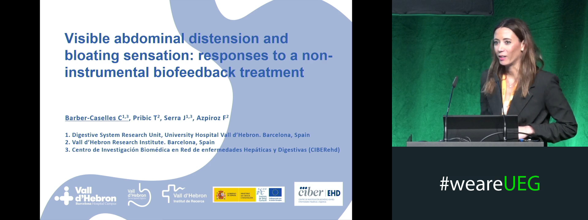
VISIBLE ABDOMINAL DISTENSION AND BLOATING SENSATION: RESPONSES TO A NON-INSTRUMENTAL BIOFEEDBACK TREATMENT.
1 University Hospital Vall d'Hebron, Barcelona, Spain
2 Vall D'Hebron Research Institute, Barcelona, Spain
Event
Submission format
Session
Citation
Published
Log in to access this content.
Free for all myUEG account holders. Your access level is set automatically based on your occupation. Medical professionals get full access to all content. If you are a non-medical user, you can only access UEG Week content from congresses you attended.
Not sure what you can access? Learn more about account types.
Should we give probiotics a chance? For what?
1 Center for Biomarker Research in Medicine (CBmed), Graz, Austria
Event
Topics
Citation
Published
Log in to access this content.
Free for all myUEG account holders. Your access level is set automatically based on your occupation. Medical professionals get full access to all content. If you are a non-medical user, you can only access UEG Week content from congresses you attended.
Not sure what you can access? Learn more about account types.
EFFECTIVENESS OF A LOW HISTAMINE DIET IN PATIENTS WITH IRRITABLE BOWEL SYNDROME: A CLINICAL STUDY
1 Clinic of Internal Diseases, Naval Hospital – Varna, Military Medical Academy, Bulgaria, Varna, Bulgaria
2 Medical University of Varna, Varna, Bulgaria
3 Medical University Varna, Varna, Bulgaria
Conference
Topics
Submission format
Session
Citation
Published
Log in to access this content.
Free for all myUEG account holders. Your access level is set automatically based on your occupation. Medical professionals get full access to all content. If you are a non-medical user, you can only access UEG Week content from congresses you attended.
Not sure what you can access? Learn more about account types.
Summary: UEG Journal Session
1 University Medical Center Maribor, Maribor, Slovenia
Event
Topics
Citation
Published

Log in to access this content.
Free for all myUEG account holders. Your access level is set automatically based on your occupation. Medical professionals get full access to all content. If you are a non-medical user, you can only access UEG Week content from congresses you attended.
Not sure what you can access? Learn more about account types.
INCIDENCE, PREVALENCE AND OVERALL RISK OF ESOPHAGEAL CANCER IN ACHALASIA: A PROPENSITY-MATCHED POPULATION-BASED STUDY FROM A LARGE MULTICENTER DATABASE
Alberto Barchi 1, Edoardo Vespa 1, Francesco Vito Mandarino 1, Antonio Ventimiglia 1, Marina Girelli 1, Micaela Marchisotta 1, Giuseppe Dell'Anna 1, Milli Gupta 2, Francesca Rusconi 3, Vito Annese 4, Edoardo Vincenzo Savarino 5, Silvio Danese 1, Sandro Passaretti 1, Albert J. Bredenoord 6
1 IRCCS Ospedale San Raffaele, Milan, Italy
2 University of Calgary, Calgary, AB, Canada
3 TriNetX Europe NV, Sint-Martens-Latem, Belgium
4 IRCCS Policlinico San Donato, Milan, Italy
5 University of Padua, Division of Gastroenterology, Padua, Italy
6 Amsterdam UMC, Amsterdam, Netherlands