UEG's content platform for gastroenterology education bringing recordings, courses, guidelines, and more together in one place.
Trending on Gutflix
Rate for better recommendations!
Your feedback helps us show you more of what you like and less of what you dislike!
Log in to continue.
This content is part of Gutflix. Log in with your myUEG account, or create one free, to watch it.
Not sure what you can access? Learn more about account types.
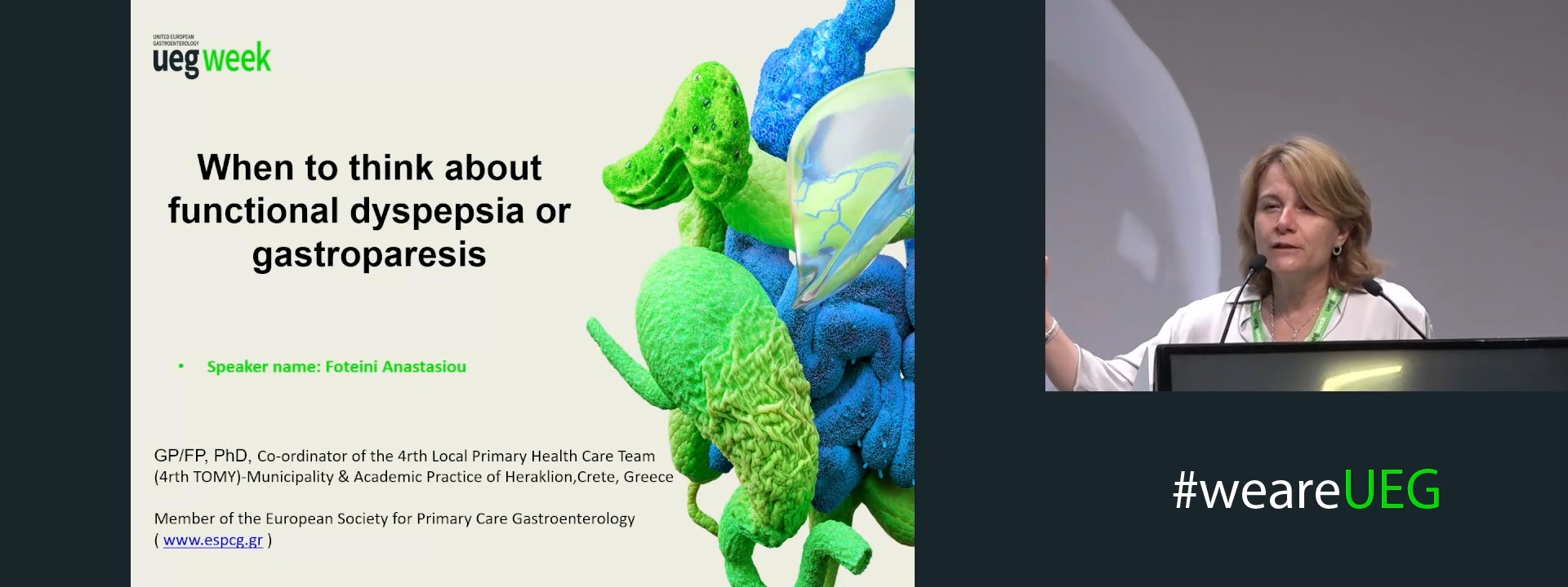
When to think about functional dyspepsia or gastroparesis
Affiliations
1 4rth Local Primary Health Care Team (TOMY) - Municipality Practice-Academic Practice of Heraklion, Crete, Heraklion, Crete, Greece
Log in to continue.
This content is part of Gutflix. Log in with your myUEG account, or create one free, to watch it.
Not sure what you can access? Learn more about account types.
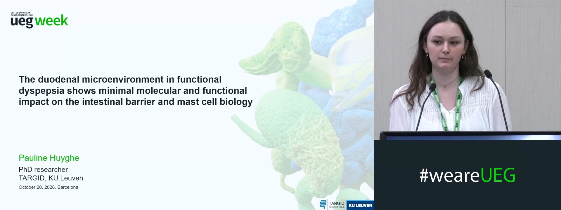
THE DUODENAL MICROENVIRONMENT IN FUNCTIONAL DYSPEPSIA SHOWS MINIMAL MOLECULAR AND FUNCTIONAL IMPACT ON THE INTESTINAL BARRIER AND MAST CELL BIOLOGY
Pauline Huyghe 1, Matthias Ceulemans 2, Theo Thijs 1, Joran Toth 1, Mona Farhadipour 3, Lorenzo Giorio 1, Kaline Arnauts 1, Åsa V. Keita 4, Bram Verstockt 5, Lucas Wauters 5, Inge Depoortere 1, Jan Tack 5, Tim Vanuytsel 5
Affiliations
1 KU Leuven, Translational Research Center for Gastrointestinal Disorders (TARGID), Leuven, Belgium
2 Precision Immunology Institute, Icahn School of Medicine at Mount Sinai, New York, United States
3 Division of Gastroenterology and Hepatology, Mayo Clinic, Rochester, United States
4 Linköping University Clinical and Exper. Medicine Faculty of Health Sciences, Linköping, Sweden
5 University Hospitals Leuven and KU Leuven, Translational Research in Gastrointestinal Disorders, Leuven, Belgium
Log in to continue.
This content is part of Gutflix. Log in with your myUEG account, or create one free, to watch it.
Not sure what you can access? Learn more about account types.
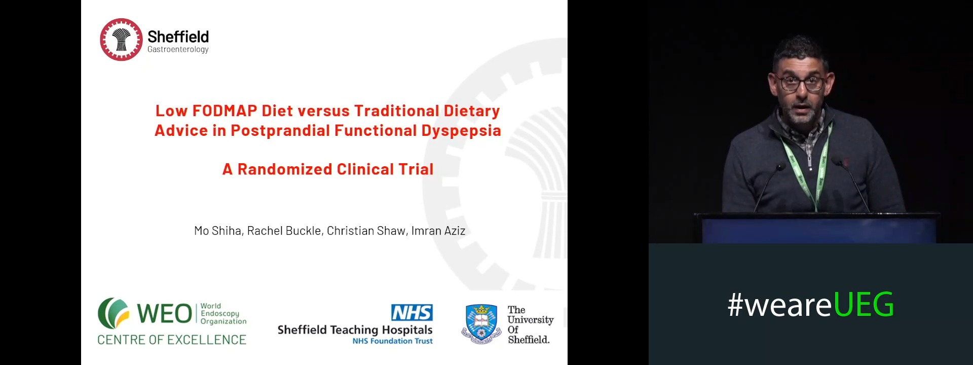
LOW FODMAP DIET VERSUS TRADITIONAL DIETARY ADVICE IN POSTPRANDIAL FUNCTIONAL DYSPEPSIA: A RANDOMIZED CLINICAL TRIAL
Affiliations
1 Division of Clinical Medicine, School of Medicine and Population Health, University of Sheffield, Sheffield, United Kingdom
2 Department of Dietetics, Sheffield Teaching Hospitals, Sheffield, United Kingdom
Log in to continue.
This content is part of Gutflix. Log in with your myUEG account, or create one free, to watch it.
Not sure what you can access? Learn more about account types.
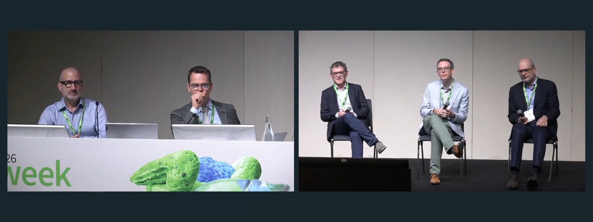
Panel discussion: Cross-cutting: Diagnostic - invasive or not invasive