UEG's educational platform for gastroenterology education bringing recordings, courses, guidelines, and more together in one place.
Most popular content
Rate for better recommendations!
Your feedback helps us show you more of what you like and less of what you dislike!
Log in to access this content.
Free for all myUEG account holders. Your access level is set automatically based on your occupation. Medical professionals get full access to all content. If you are a non-medical user, you can only access UEG Week content from congresses you attended.
Not sure what you can access? Learn more about account types.

GUSELKUMAB VERSUS RISANKIZUMAB AS MAINTENANCE TREATMENT FOR MODERATELY TO SEVERELY ACTIVE ULCERATIVE COLITIS: NETWORK META-ANALYSES OF CLINICAL REMISSION AND ENDOSCOPIC OUTCOMES
1 Johnson & Johnson, Horsham, PA, United States
2 Janssen Pharmaceutica, Beerse, Belgium
3 Johnson & Johnson, Spring House, PA, United States
4 EVERSANA Value & Evidence Services, Burlington, ON, Canada
Conference
Topics
Submission format
Citation
Published
Log in to access this content.
Free for all myUEG account holders. Your access level is set automatically based on your occupation. Medical professionals get full access to all content. If you are a non-medical user, you can only access UEG Week content from congresses you attended.
Not sure what you can access? Learn more about account types.
Innovations in IBD Therapy: The Story of TL1A - from Discovery to Potential Future Care (F. Hoffmann-La Roche Ltd) (Complete Session)
1 Icahn School of Medicine at Mount Sinai, USA, United States of America
2 Hospital Universitario Reina Sofia, Cordoba, Spain
3 University of Calgary, Calgary, Canada
Publisher
Event
Citation
Published
Log in to access this content.
Free for all myUEG account holders. Your access level is set automatically based on your occupation. Medical professionals get full access to all content. If you are a non-medical user, you can only access UEG Week content from congresses you attended.
Not sure what you can access? Learn more about account types.
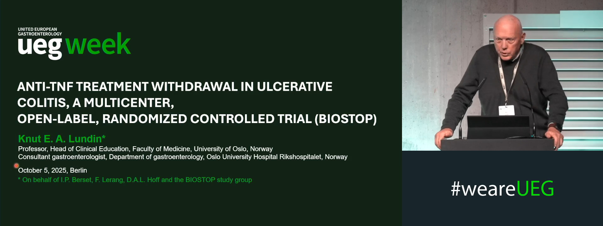
ANTI-TNF TREATMENT WITHDRAWAL IN ULCERATIVE COLITIS, A MULTICENTER, OPEN-LABEL, RANDOMIZED CONTROLLED TRIAL
1 Gastroenterology Aalesund, Alesund, Norway
2 Central Hosspital of Ostfold, Grålum, Norway
3 Aalesund Hospital, Møre & Romsdal Hospital trust, Aalesund, Norway
4 K. G. Jebsen Coeliac Disease Research Centre, University of Oslo and Oslo University Hospital, Oslo, Norway
Event
Submission format
Session
Citation
Published
Log in to access this content.
Free for all myUEG account holders. Your access level is set automatically based on your occupation. Medical professionals get full access to all content. If you are a non-medical user, you can only access UEG Week content from congresses you attended.
Not sure what you can access? Learn more about account types.
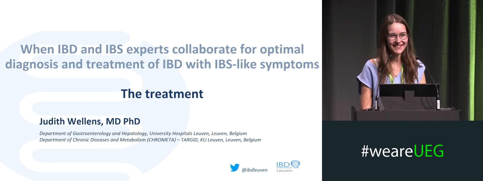
The treatment
1 University Hospitals Leuven, Leuven, Belgium
Event
Topics
Citation
Published
Log in to access this content.
Free for all myUEG account holders. Your access level is set automatically based on your occupation. Medical professionals get full access to all content. If you are a non-medical user, you can only access UEG Week content from congresses you attended.
Not sure what you can access? Learn more about account types.
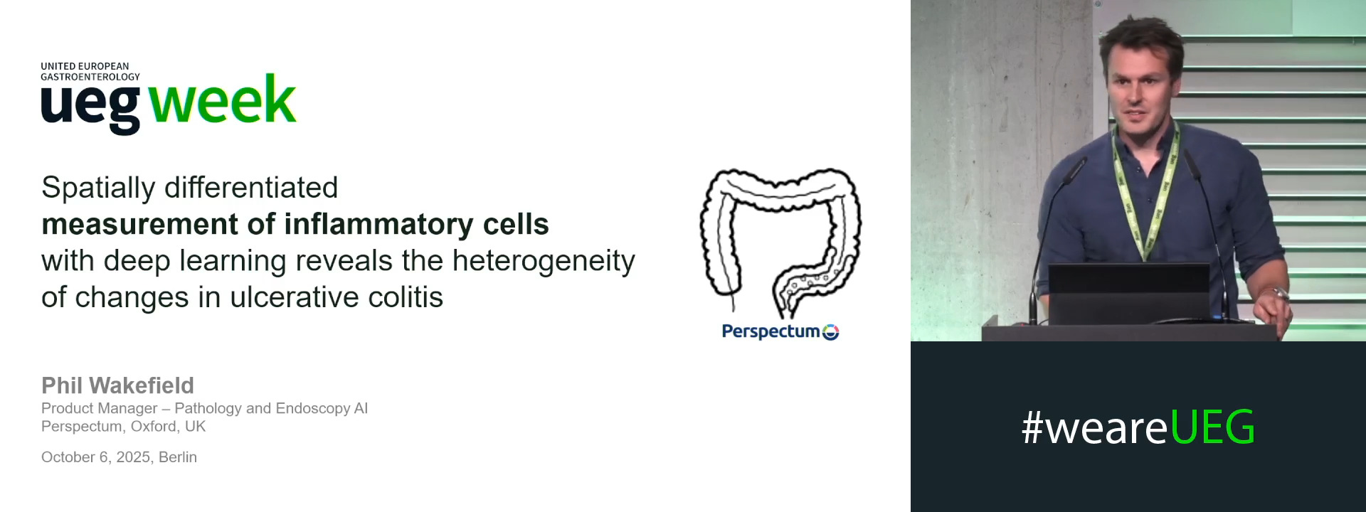
SPATIALLY DIFFERENTIATED MEASUREMENT OF INFLAMMATORY CELLS WITH DEEP LEARNING REVEALS THE HETEROGENEITY OF CHANGES IN ULCERATIVE COLITIS
Dylan Windell 1, Alastair Magness 1, Rongqi Li 1, Paul Aljabar 1, Eve Fryer 2, Robert D. Goldin 3, Phil Wakefield 1, Caitlin Langford 4, Jonathan Landy 5
1 Perspectum Ltd., Oxford, United Kingdom
2 John Radcliffe Hospital, Oxford, United Kingdom
3 Centre for Pathology, London, United Kingdom
4 Perspectum, Cambridge, United States
5 London North West University Healthcare NHS Trust, London, United Kingdom
Event
Topics
Submission format
Session
Citation
Published
Log in to access this content.
Free for all myUEG account holders. Your access level is set automatically based on your occupation. Medical professionals get full access to all content. If you are a non-medical user, you can only access UEG Week content from congresses you attended.
Not sure what you can access? Learn more about account types.
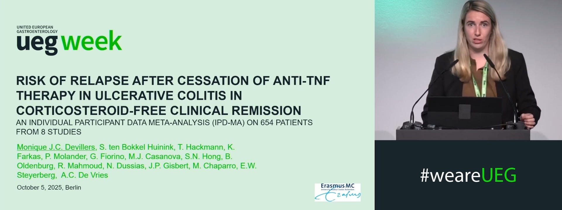
RISK OF RELAPSE AFTER CESSATION OF ANTI-TNF THERAPY IN ULCERATIVE COLITIS IN CORTICOSTEROID-FREE CLINICAL REMISSION: AN INDIVIDUAL PARTICIPANT DATA META-ANALYSIS (IPD-MA) ON 654 PATIENTS FROM 8 STUDIES
Monique Devillers 1, Sebastiaan ten Bokkel Huinink 1, Toby Hackmann 2, Ewout Steyerberg 3, Bas Oldenburg 4, Remi Mahmoud 4, Pauliina Molander 5, Klaudia Farkas 6, Gionata Fiorino 7, María Chaparro 8, Javier Gisbert 8, María José Casanova 8, Sung-Noh Hong 9, Nikolas Dussias 10, Annemarie de Vries 1
1 Erasmus MC University Medical Center, Rotterdam, Netherlands
2 Leiden University Medical Center, Leiden, Netherlands
3 Utrecht University Medical Center, Utrecht, Netherlands
4 University Medical Center Utrecht, Utrecht, Netherlands
5 Helsinki University Hospital, Helsinki, Finland
6 University of Szeged 1st Dept. of Medicine, Szeged, Hungary
7 San Camillo-Forlanini Hospital, Rome, Italy
8 Hospital Universitario de La Princesa, Instituto de Investigación Sanitaria Princesa (IIS-Princesa), Universidad Autónoma de Madrid (UAM), Centro de Investigación Biomédica en Red de Enfermedades Hepáticas y Digestivas (CIBEREHD), Madrid, Spain
9 Samsung Medical Center, Seoul, Korea, Republic of
10 IRCCS Azienda Ospedaliero-Universitaria di Bologna, Bologna, Italy
Event
Submission format
Session
Citation
Published
Log in to access this content.
Free for all myUEG account holders. Your access level is set automatically based on your occupation. Medical professionals get full access to all content. If you are a non-medical user, you can only access UEG Week content from congresses you attended.
Not sure what you can access? Learn more about account types.
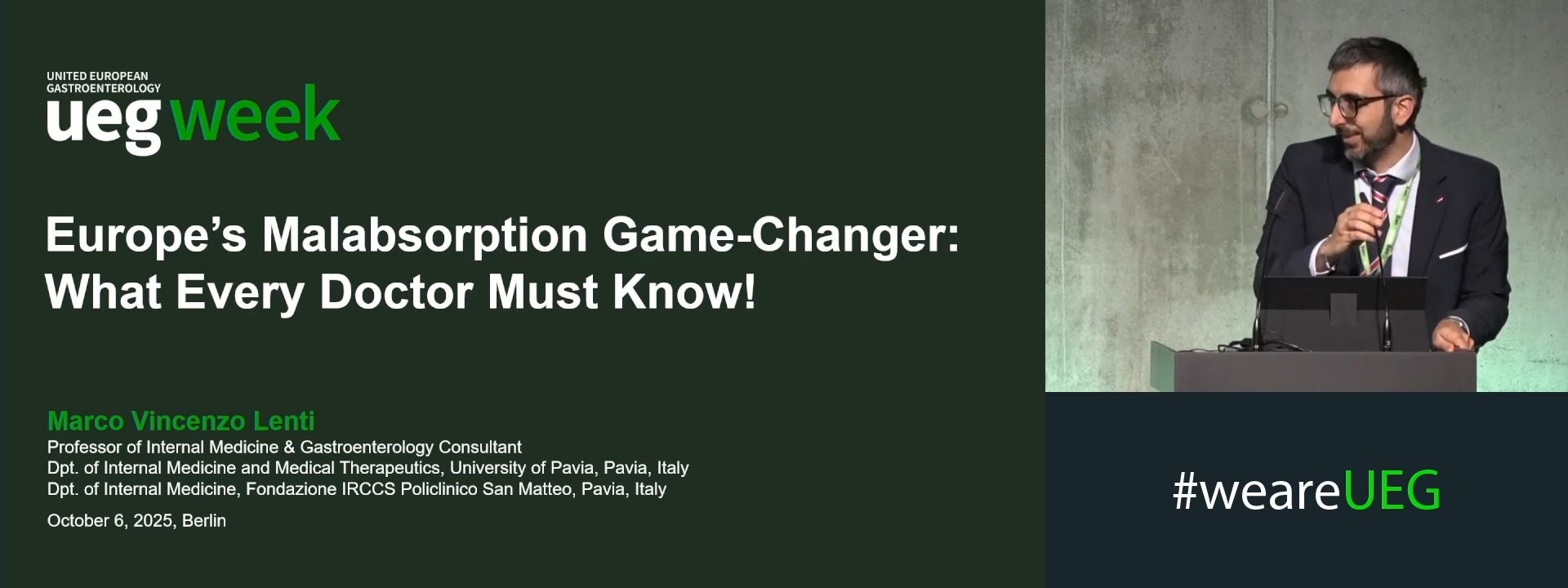
Europe’s malabsorption game-changer: What every doctor must know!
1 Fondazione IRCCS Policlinico San Matteo, University of Pavia, Alessandria, Italy