UEG's educational platform for gastroenterology education bringing recordings, courses, guidelines, and more together in one place.
Trending on Gutflix
Rate for better recommendations!
Your feedback helps us show you more of what you like and less of what you dislike!
Log in to continue.
This content is part of Gutflix. Log in with your myUEG account, or create one free, to watch it.
Not sure what you can access? Learn more about account types.
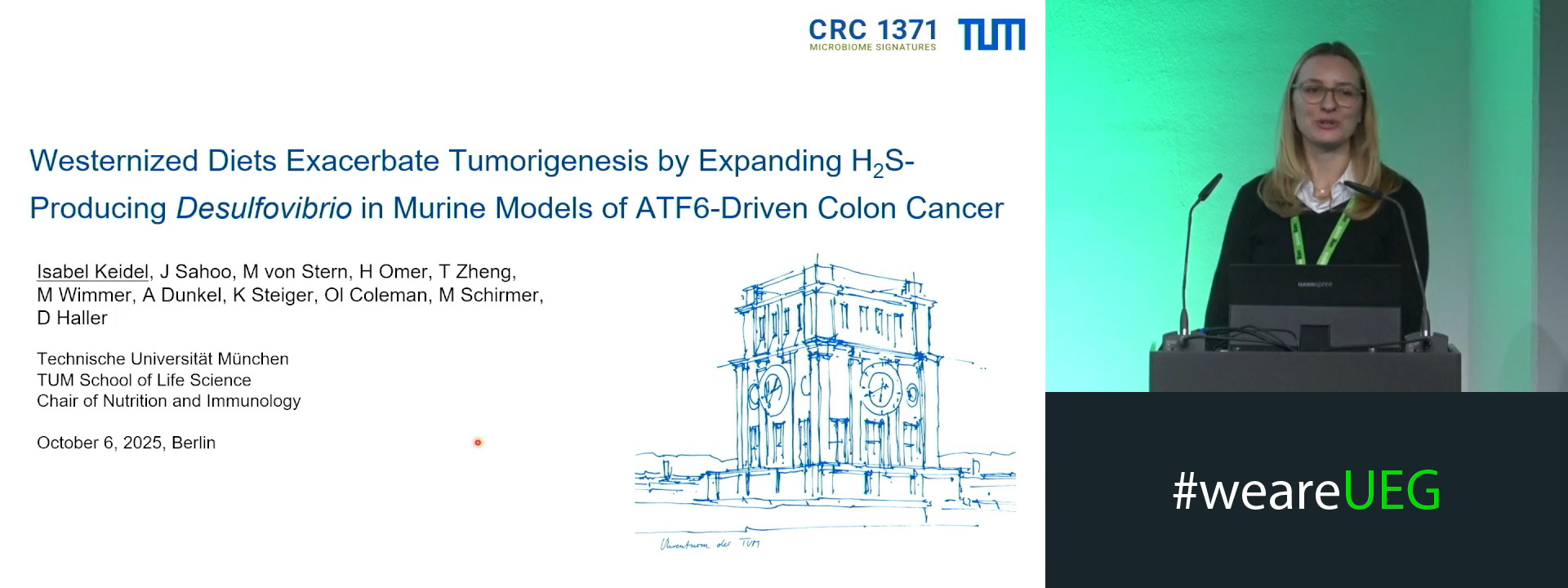
WESTERNIZED DIETS EXACERBATE TUMORIGENESIS BY EXPANDING H2S-PRODUCING DESULFOVIBRIO IN MURINE MODELS OF ATF6-DRIVEN COLON CANCER
Isabel Keidel 1, Jagadrupa Sahoo 1, Miriam von Stern-Gwiazdowski 2, Hélène Omer 1, Tingting Zheng 1, Miriam Wimmer 2, Andreas Dunkel 2, Katja Steiger 3, Olivia Coleman 2, Melanie Schirmer 1, Dirk Haller 1
Affiliations
1 Technical University of Munich, Freising, Germany|||Technical University of Munich, Freising, Germany
2 Technical University of Munich, Freising, Germany
3 Technical University of Munich, München, Germany
Log in to continue.
This content is part of Gutflix. Log in with your myUEG account, or create one free, to watch it.
Not sure what you can access? Learn more about account types.
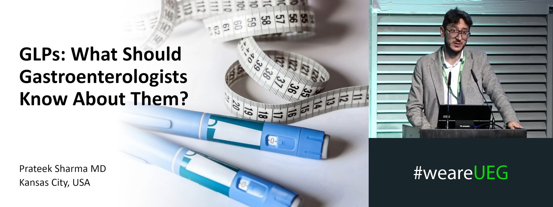
GLPs: What should gastroenterologists know about them?
Affiliations
1 University of Kansas School of Medicine Dept. of Gastroenterology, Leawood, United States of America
Log in to continue.
This content is part of Gutflix. Log in with your myUEG account, or create one free, to watch it.
Not sure what you can access? Learn more about account types.
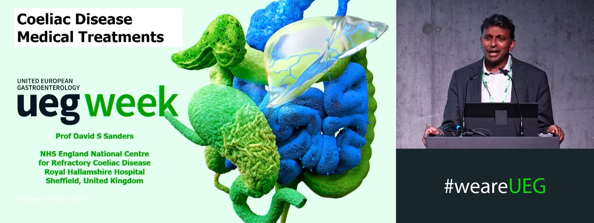
Medical treatment
Affiliations
1 Royal Hallamshire Hospital - NHS Trust, Royal Hallamshire Hospital; Sheffield/GB, Sheffield, United Kingdom
Log in to continue.
This content is part of Gutflix. Log in with your myUEG account, or create one free, to watch it.
Not sure what you can access? Learn more about account types.
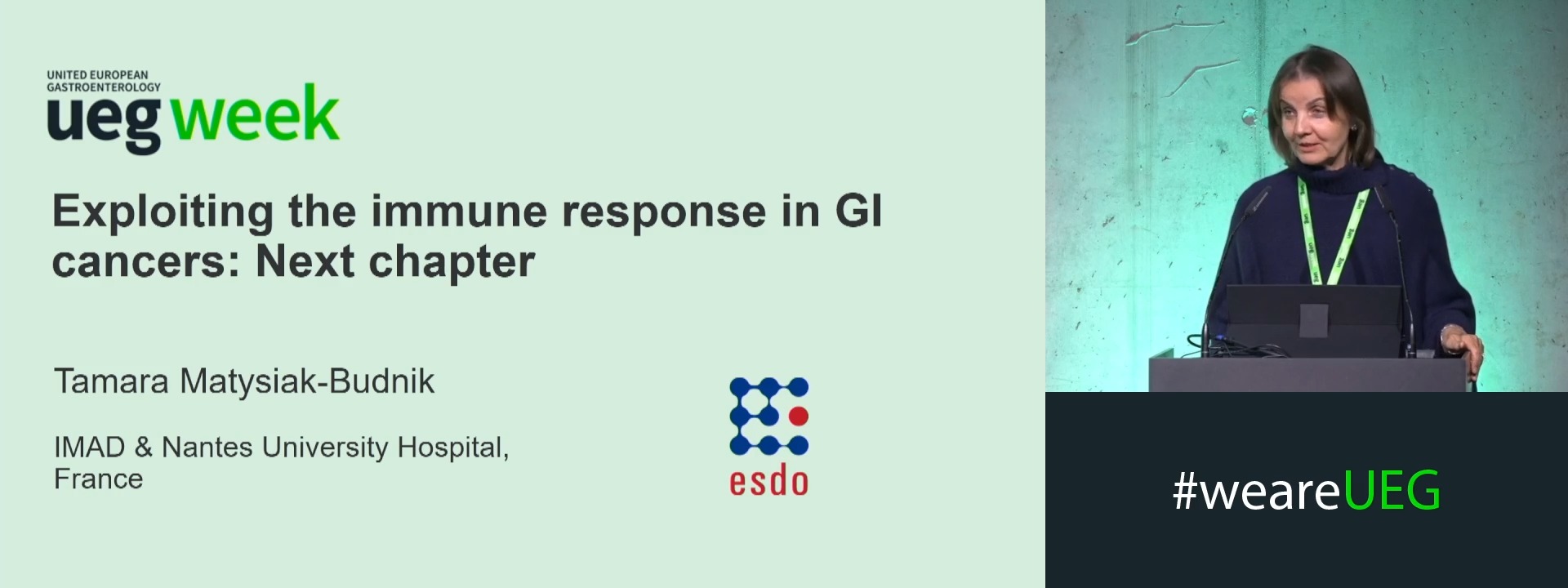
Exploiting the immune response in GI cancer: Next chapter
Affiliations
1 CHU de Nantes IMAD, Nantes, France
Log in to continue.
This content is part of Gutflix. Log in with your myUEG account, or create one free, to watch it.
Not sure what you can access? Learn more about account types.
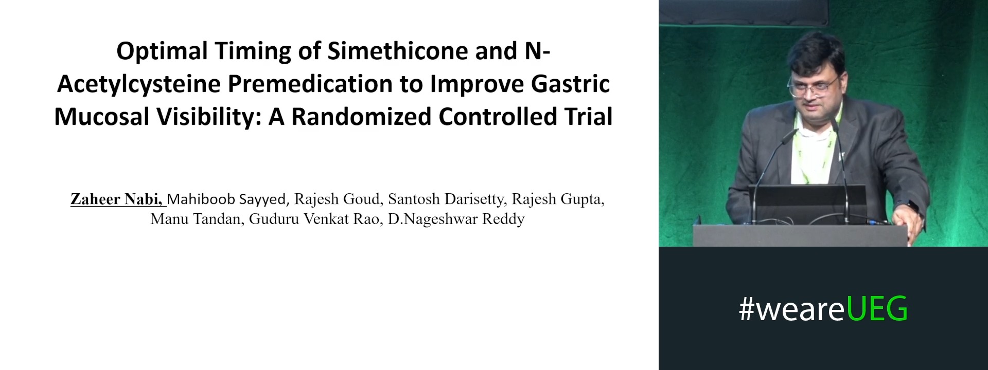
IMPACT OF TIMING OF PREMEDICATION ON MUCOSAL VISIBILITY DURING ENDOSCOPY-A RANDOMIZED CONTROLLED TRIAL
Affiliations
1 Asian Institute of Gastroenterology, Hyderabad, India
2 Asian Institute of gastroenterology,Hyderabad, Hyderabad, India
Log in to continue.
This content is part of Gutflix. Log in with your myUEG account, or create one free, to watch it.
Not sure what you can access? Learn more about account types.
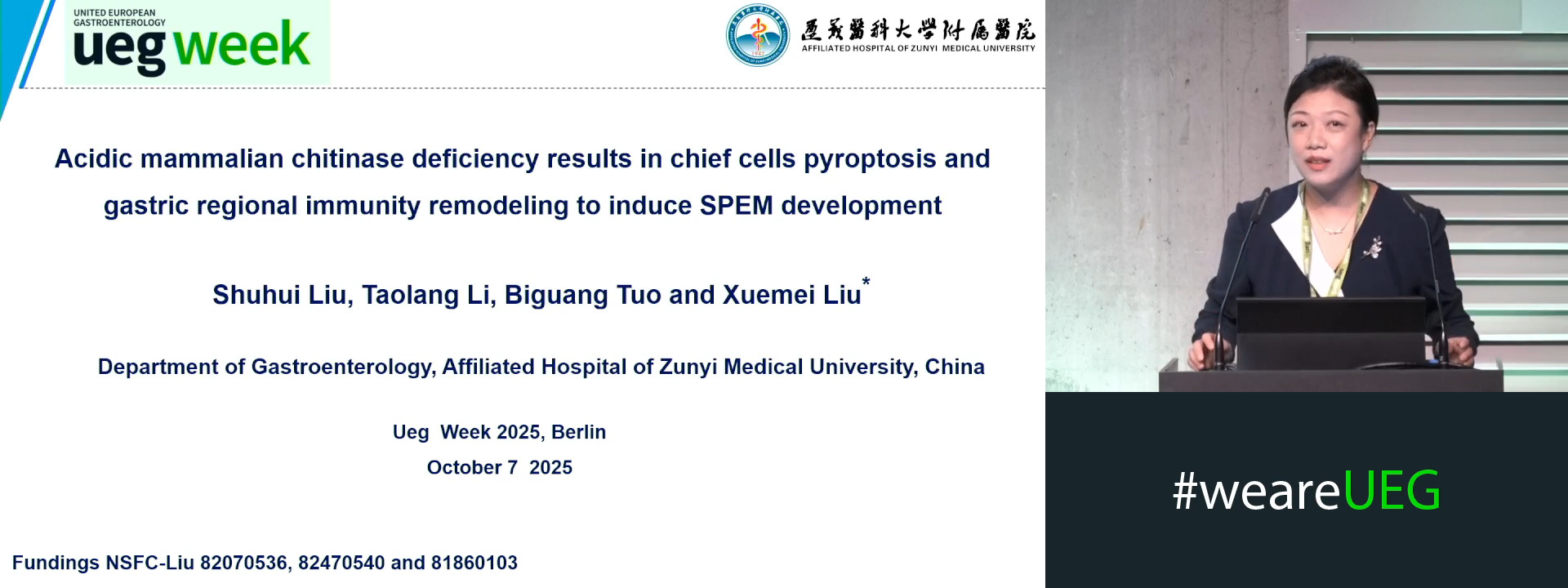
ACIDIC MAMMALIAN CHITINASE DEFICIENCY RESULTS IN CHIEF CELLS PYROPTOSIS AND GASTRIC REGIONAL IMMUNITY REMODELING IS THE NEW CELLULAR ORIGIN OF SPEM
Affiliations
1 Affiliated Hospital of Zunyi Medical University, Zunyi, China
Log in to continue.
This content is part of Gutflix. Log in with your myUEG account, or create one free, to watch it.
Not sure what you can access? Learn more about account types.
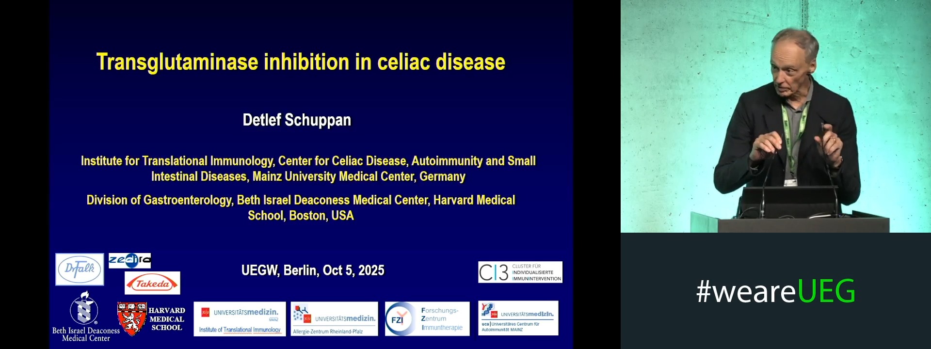
Transglutaminase inhibition in CeD
Affiliations
1 Medical University Mainz - Institute of Translational Immunology, Medical University Mainz ; Mainz/D, Mainz, Germany