UEG's content platform for gastroenterology education bringing recordings, courses, guidelines, and more together in one place.
Trending on Gutflix
Rate for better recommendations!
Your feedback helps us show you more of what you like and less of what you dislike!
Log in to continue.
This content is part of Gutflix. Log in with your myUEG account, or create one free, to watch it.
Not sure what you can access? Learn more about account types.
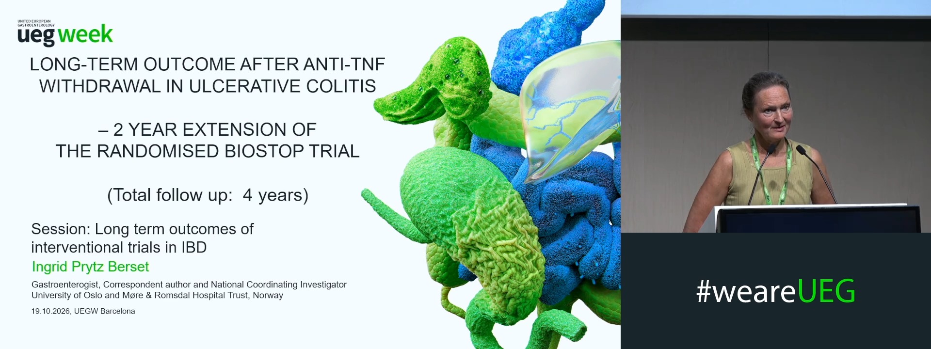
LONG-TERM OUTCOME AFTER ANTI-TNF WITHDRAWAL IN ULCERATIVE COLITIS – 2 YEAR EXTENSION OF THE RANDOMISED BIOSTOP STUDY
Ingrid Prytz Berset 1, Knut E.A. Lundin 2, Dag Arne Lihaug Hoff 3, Frode Lerang 4, Tor Åge Myklebust 5
Affiliations
1 Gastroenterology Aalesund, Alesund, Norway
2 Norwegian Coeliac Disease Research Centre, University of Oslo and Oslo University Hospital, Oslo, Norway
3 Aalesund Hospital, Møre & Romsdal Hospital trust, Aalesund, Norway
4 Central Hospital of Ostfold, Grålum, Norway
5 Aalesund Hospital, Møre & Romsdal Hospital Trust, Aalesund, Norway
Log in to continue.
This content is part of Gutflix. Log in with your myUEG account, or create one free, to watch it.
Not sure what you can access? Learn more about account types.
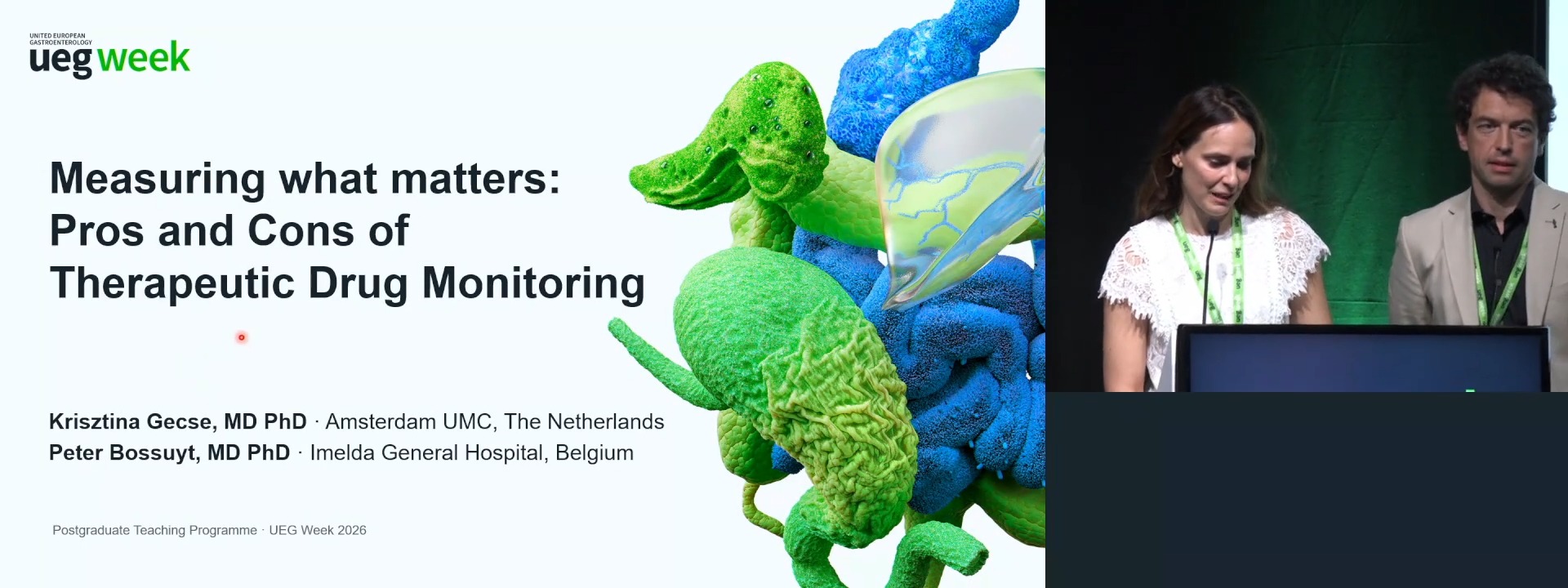
Measuring what matters: Pros and cons of therapeutic drug monitoring in IBD
Affiliations
1 Department of Gastroenterology and Hepatology, Amsterdam, Netherlands
2 Imelda General Hospital, Bonheiden, Belgium
Log in to continue.
This content is part of Gutflix. Log in with your myUEG account, or create one free, to watch it.
Not sure what you can access? Learn more about account types.
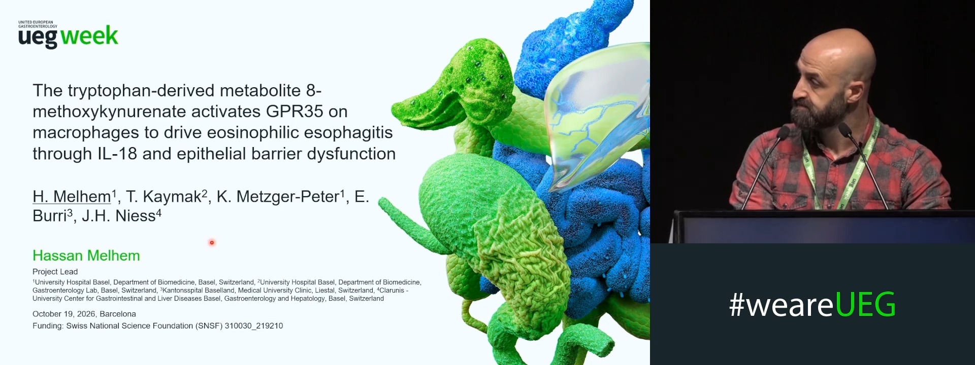
THE TRYPTOPHAN-DERIVED METABOLITE 8-METHOXYKYNURENATE ACTIVATES GPR35 ON MACROPHAGES TO DRIVE EOSINOPHILIC ESOPHAGITIS THROUGH IL-18 AND EPITHELIAL BARRIER DYSFUNCTION
Affiliations
1 University Hospital Basel, Basel, Switzerland
2 Kantonsspital Baselland, Liestal, Switzerland
3 Clarunis - University Center for Gastrointestinal and Liver Diseases Basel, Basel, Switzerland
Log in to continue.
This content is part of Gutflix. Log in with your myUEG account, or create one free, to watch it.
Not sure what you can access? Learn more about account types.
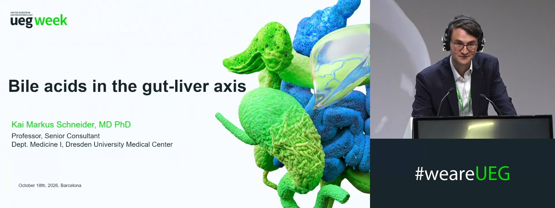
Bile acids in the gut-liver axis
Affiliations
1 Dresden University Medical Center, Dresden, Germany
Log in to continue.
This content is part of Gutflix. Log in with your myUEG account, or create one free, to watch it.
Not sure what you can access? Learn more about account types.
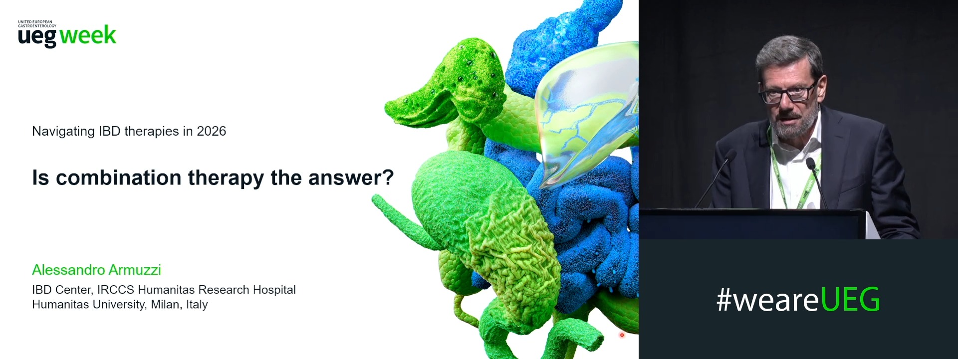
Is combination therapy the answer?
Affiliations
1 IBD Center, IRCCS Humanitas Research Hospital, Rozzano, Milan, Italy
Log in to continue.
This content is part of Gutflix. Log in with your myUEG account, or create one free, to watch it.
Not sure what you can access? Learn more about account types.
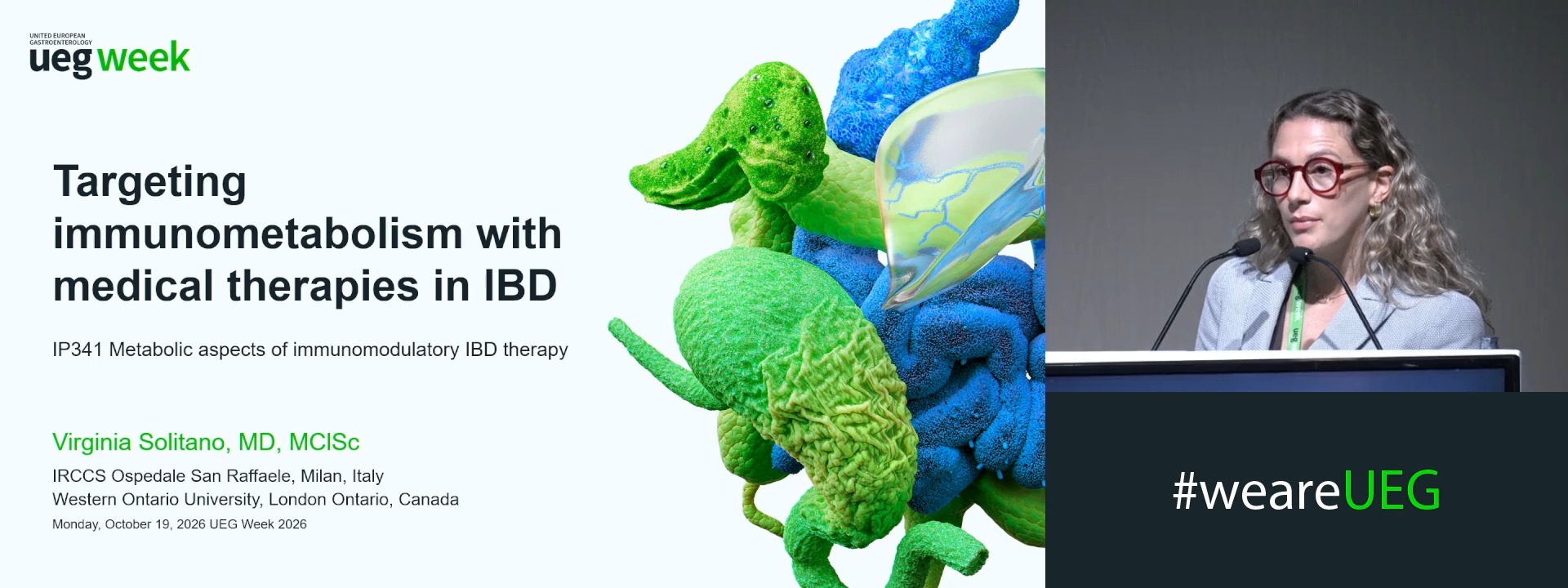
Targeting immunometabolism with medical therapies in IBD
Affiliations
1 IRCCS Ospedale San Raffaele, Milan, Italy
Log in to continue.
This content is part of Gutflix. Log in with your myUEG account, or create one free, to watch it.
Not sure what you can access? Learn more about account types.
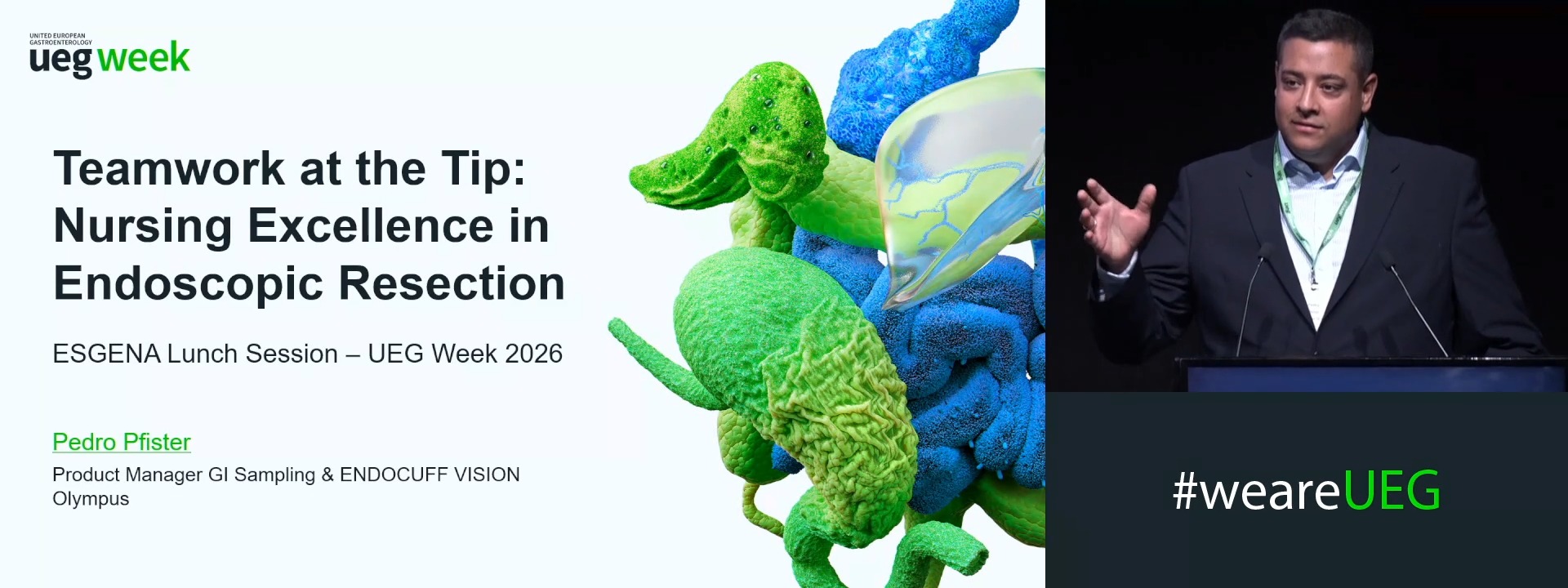
Teamwork at the tip: Nursing excellence in endoscopic resection (OLYMPUS EUROPA SE & CO. KG )