UEG's educational platform for gastroenterology education bringing recordings, courses, guidelines, and more together in one place.
Most popular content
Rate for better recommendations!
Your feedback helps us show you more of what you like and less of what you dislike!
Log in to access this content.
Free for all myUEG account holders. Your access level is set automatically based on your occupation. Medical professionals get full access to all content. If you are a non-medical user, you can only access UEG Week content from congresses you attended.
Not sure what you can access? Learn more about account types.
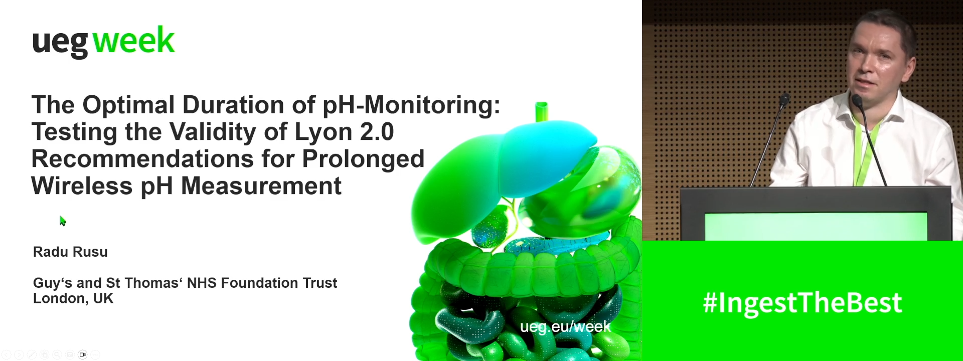
THE OPTIMAL DURATION OF PH-MONITORING: TESTING THE VALIDITY OF LYON 2.0 RECOMMENDATIONS FOR PROLONGED WIRELESS PH MEASUREMENT
Radu-Ionut Rusu 1, Mark Fox 2, Rami Sweis 3, Sebastian S. Zeki 1, Jason M. Dunn 1, Angela Anggiansah 1, Jafar Jafari 1, Annastazia Learoyd 4, Terence Wong 1
1 Guy's and St Thomas' NHS Foundation Trust, London, United Kingdom
2 Klinik Arlesheim & Zürich University, Arlesheim, Switzerland
3 University College Hospital, London, United Kingdom
4 Faculty of Life Sciences & Medicine, King's College London, London, United Kingdom
Event
Submission format
Session
Citation
Published
Log in to access this content.
Free for all myUEG account holders. Your access level is set automatically based on your occupation. Medical professionals get full access to all content. If you are a non-medical user, you can only access UEG Week content from congresses you attended.
Not sure what you can access? Learn more about account types.
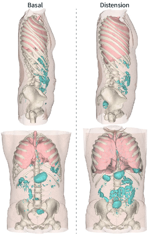
Mistakes in abdominal distension and how to avoid them
Citation
Published
Log in to access this content.
Free for all myUEG account holders. Your access level is set automatically based on your occupation. Medical professionals get full access to all content. If you are a non-medical user, you can only access UEG Week content from congresses you attended.
Not sure what you can access? Learn more about account types.
EoE with Alex Straumann - Part 2
Topics
Published
Log in to access this content.
Free for all myUEG account holders. Your access level is set automatically based on your occupation. Medical professionals get full access to all content. If you are a non-medical user, you can only access UEG Week content from congresses you attended.
Not sure what you can access? Learn more about account types.

Esophageal and Oropharyngeal Dysphagia: Clinical Recommendations From the United European Gastroenterology and European Society for Neurogastroenterology and Motility
Publisher
Guideline
Citation
Published
Log in to access this content.
Free for all myUEG account holders. Your access level is set automatically based on your occupation. Medical professionals get full access to all content. If you are a non-medical user, you can only access UEG Week content from congresses you attended.
Not sure what you can access? Learn more about account types.

European Guideline on Pre-Operative Prevention of Surgical Site Infections Following Digestive Surgery: A Joint Update of the WHO SSI Guideline for Gastrointestinal Surgery by UEG, ESCP, EAES, and SIS-E
Publishers
Guideline
Topics
Citation
Published
Log in to access this content.
Free for all myUEG account holders. Your access level is set automatically based on your occupation. Medical professionals get full access to all content. If you are a non-medical user, you can only access UEG Week content from congresses you attended.
Not sure what you can access? Learn more about account types.
Mistakes in rumination syndrome and how to avoid them
Citation
Published
Log in to access this content.
Free for all myUEG account holders. Your access level is set automatically based on your occupation. Medical professionals get full access to all content. If you are a non-medical user, you can only access UEG Week content from congresses you attended.
Not sure what you can access? Learn more about account types.