UEG's educational platform for gastroenterology education bringing recordings, courses, guidelines, and more together in one place.
Most popular content
Rate for better recommendations!
Your feedback helps us show you more of what you like and less of what you dislike!
Log in to access this content.
Free for all myUEG account holders. Your access level is set automatically based on your occupation. Medical professionals get full access to all content. If you are a non-medical user, you can only access UEG Week content from congresses you attended.
Not sure what you can access? Learn more about account types.

PREDICTION OF PANCREATIC NEUROENDOCRINE TUMOR GRADING USING AN ARTIFICIAL INTELLIGENCE-BASED VIDEO ANALYSIS MODEL APPLIED TO CONTRAST-ENHANCED ENDOSCOPIC ULTRASOUND VIDEOS
Matteo Tacelli 1, Adrien Meyer 2, Paolo Biamonte 1, Armine Vardazaryan 2, Rubino Nunziata 1, Leonardo Sosa Valencia 2, Gabriele Capurso 1, Nicolas Padoy 2, Paolo Giorgio Arcidiacono 1
1 San Raffaele Scientific Institute IRCCS, Milano, Italy
2 IHU-Strasbourg, Institute of Image-Guided Surgery, Strasbourg, France
Conference
Submission format
Session
Citation
Published
Log in to access this content.
Free for all myUEG account holders. Your access level is set automatically based on your occupation. Medical professionals get full access to all content. If you are a non-medical user, you can only access UEG Week content from congresses you attended.
Not sure what you can access? Learn more about account types.
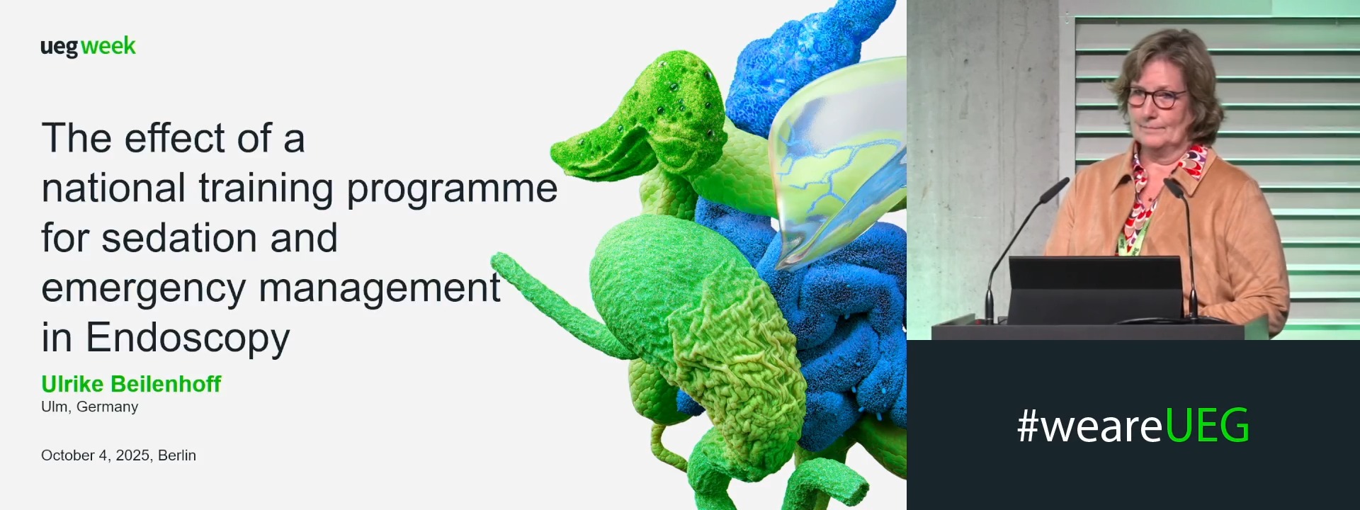
THE EFFECT OF A NATIONAL TRAINING PROGRAMME FOR SEDATION AND EMERGENCY MANAGEMENT IN ENDOSCOPY
1 ESGENA Scientific Secretary, Ulm, Germany
Event
Topics
Submission format
Citation
Published
Log in to access this content.
Free for all myUEG account holders. Your access level is set automatically based on your occupation. Medical professionals get full access to all content. If you are a non-medical user, you can only access UEG Week content from congresses you attended.
Not sure what you can access? Learn more about account types.
USEFULNESS OF LYMPHOCYTE/MONOCYTE RATIO IN PREDICTING PROGNOSIS IN PATIENTS RECEIVING ATEZOLIZUMAB + BEVACIZUMAB
Emi Tanaka 1, Toshifumi Tada 2, HANA OTA 1, Kaho Aoe 1, Atsushi Hiraoka 3, Hideko Ohama 3, Hidenori Toyoda 4, Kazuya Kariyama 5, Takeshi Hatanaka 6, Kazuhiro Nouso 5, Satoru Kakizaki 7, Takashi Kumada 8, Shinichiro Nakamura 1
1 Japanese Red Cross Society Himeji Hospital, Himeji, Japan
2 Kobe University Graduate School of Medicine, Kobe, Japan|||Japanese Red Cross Society Himeji Hospital, Himeji, Japan
3 Ehime Prefectural Central Hospital, Matsuyama, Japan
4 Ogaki Municipal Hospital, Ogaki, Japan
5 Okayama City Hospital, Okayama, Japan
6 Gunma Saiseikai Maebashi Hospital, Maebashi, Japan
7 NHO Takasaki General Medical Center, Takasaki, Japan
8 Gifu Kyoritsu University, Ogaki, Japan
Event
Submission format
Citation
Published
Log in to access this content.
Free for all myUEG account holders. Your access level is set automatically based on your occupation. Medical professionals get full access to all content. If you are a non-medical user, you can only access UEG Week content from congresses you attended.
Not sure what you can access? Learn more about account types.
AI-BUS: ARTIFICIAL INTELLIGENCE BOWEL ULTRASOUND SYSTEM
Jonatan Ruiz-Molsgaard 1, Jakob Karrer 1, Bobby Lo 2, Bjørn Leth Møller 3, Gorm Roager Madsen 2, Johan Fremberg Ilvemark 4, Klaus Theede 2, Johan Burisch 2, Bulat Ibragimov 3, Trine Boysen 2
1 University of copenhagen, Copenhagen, Denmark|||Hvidovre Hospital, Hvidovre, Denmark
2 Hvidovre Hospital, Hvidovre, Denmark
3 University of copenhagen, Copenhagen, Denmark
4 Herlev University Hospital, Hvidovre, Denmark
Event
Topics
Submission format
Session
Citation
Published
Log in to access this content.
Free for all myUEG account holders. Your access level is set automatically based on your occupation. Medical professionals get full access to all content. If you are a non-medical user, you can only access UEG Week content from congresses you attended.
Not sure what you can access? Learn more about account types.
GASTRIC ISCHEMIA: THE FORGOTTEN CULPRIT IN ABDOMINAL PAIN
Walid Nasreddine 1, Abbas Chamsuddin 2, Hind Joumaa 3, Fatima Al Salaman 3, Alaa Hajjar 3, Mohamad Siblini 1, Lama Chouman 4
1 Makassed General Hospital, Beirut, Lebanon
2 Saint George University of Beirut, Beirut, Lebanon
3 Beirut Arab University, Beirut, Lebanon
4 Makassed Hospital, Beirut, Lebanon
Conference
Topics
Submission format
Session
Published
Log in to access this content.
Free for all myUEG account holders. Your access level is set automatically based on your occupation. Medical professionals get full access to all content. If you are a non-medical user, you can only access UEG Week content from congresses you attended.
Not sure what you can access? Learn more about account types.
DEDICATED PRACTICE FOR PATIENTS DISCHARGED AFTER GASTROINTESTINAL BLEEDING. A SINGLE CENTER COHORT STUDY AND COST-EFFECTIVENESS ANALYSIS
Clelia Marmo 1, Daniela Feliciani 1, Barbara Funaro 1, Riccardo Marmo 2, Antonio Gasbarrini 1, Maria Elena Riccioni 1
1 Fondazione Policlinico Universitario Agostino Gemelli, IRCCS, Università Cattolica del Sacro Cuore, Rome, Italy
2 Hospital L. Curto, Polla (Salerno), Italy
Conference
Topics
Submission format
Citation
Published
Log in to access this content.
Free for all myUEG account holders. Your access level is set automatically based on your occupation. Medical professionals get full access to all content. If you are a non-medical user, you can only access UEG Week content from congresses you attended.
Not sure what you can access? Learn more about account types.
TARGETED PHAGE INTERVENTION IMPROVES BACTERIAL-DRIVEN COLITIS AND RESPONSE TO TOPICAL CORTICOSTEROIDS BY ATTENUATING VIRULENCE OF CROHN’S DISEASE-ASSOCIATED BACTERIA
Kyle Jackson 1, Heather Galipeau 2, Amber Hann 2, Marco Constante 2, Megan Zangara 3, Alexandra Fuentas 2, Chiko Shimbori 2, Premysl Bercik 2, Brian Coombes 3, Zeinab Hosseinidoust 1, Elena Verdu 2
1 McMaster University, Hamilton, Canada|||Farncombe Family Digestive Health Research Institute, Hamilton, Canada
2 Farncombe Family Digestive Health Research Institute, Hamilton, Canada
3 McMaster University, Hamilton, Canada
Event
Topics