UEG's educational platform for gastroenterology education bringing recordings, courses, guidelines, and more together in one place.
Most popular content
Rate for better recommendations!
Your feedback helps us show you more of what you like and less of what you dislike!
Log in to access this content.
Free for all myUEG account holders. Your access level is set automatically based on your occupation. Medical professionals get full access to all content. If you are a non-medical user, you can only access UEG Week content from congresses you attended.
Not sure what you can access? Learn more about account types.

LOST EXPRESSION, FOUND FAMILIES: HOW IHC REVEALS LYNCH SYNDROME BEYOND AGE AND FAMILY HISTORY
chiara montanaro 1, Filippo Facci 1, Francesca Corder 1, Riccardo Rebuzzi 1, Stefania Maiero 2, Paola Pelizzo 2, Manuela Santarosa 2, Laura Ghelfi 2, Claudio Belluco 2, Alessandra Viel 2, Renato Cannizzaro 2, Stefano Realdon 2
1 Università di Trieste, Trieste, Italy
2 Centro Riferimento Oncologico (CRO) Aviano IRCCS, Aviano, Italy
Conference
Topics
Submission format
Session
Citation
Published
Log in to access this content.
Free for all myUEG account holders. Your access level is set automatically based on your occupation. Medical professionals get full access to all content. If you are a non-medical user, you can only access UEG Week content from congresses you attended.
Not sure what you can access? Learn more about account types.
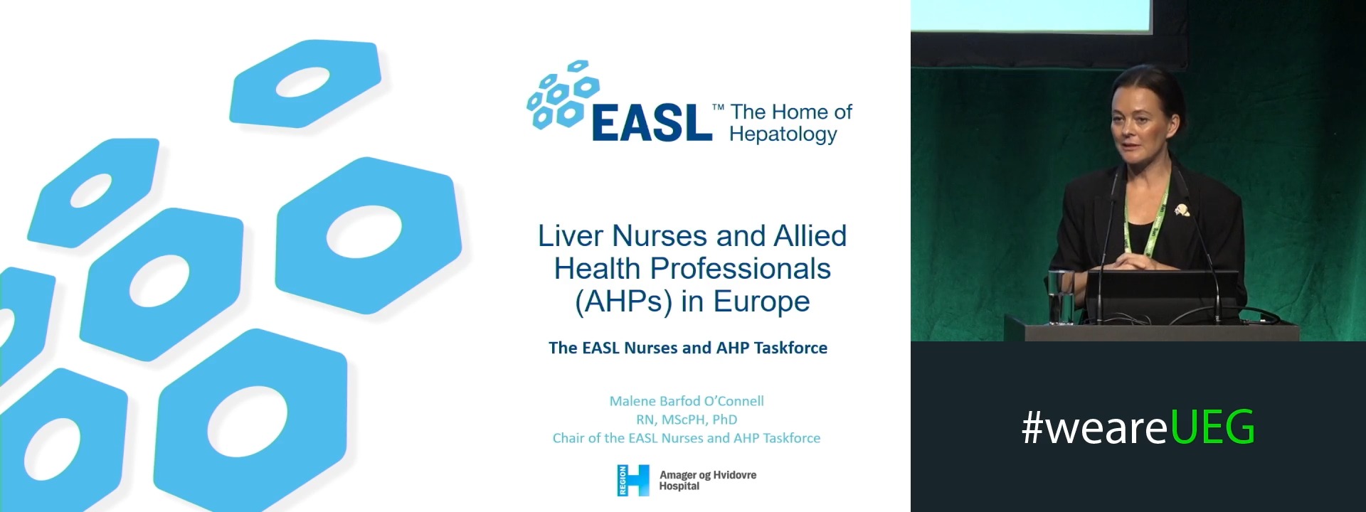
Liver nurses and allied health professions in Europe: The EASL taskforce
1 None, None, Denmark
Event
Topics
Citation
Published
Log in to access this content.
Free for all myUEG account holders. Your access level is set automatically based on your occupation. Medical professionals get full access to all content. If you are a non-medical user, you can only access UEG Week content from congresses you attended.
Not sure what you can access? Learn more about account types.
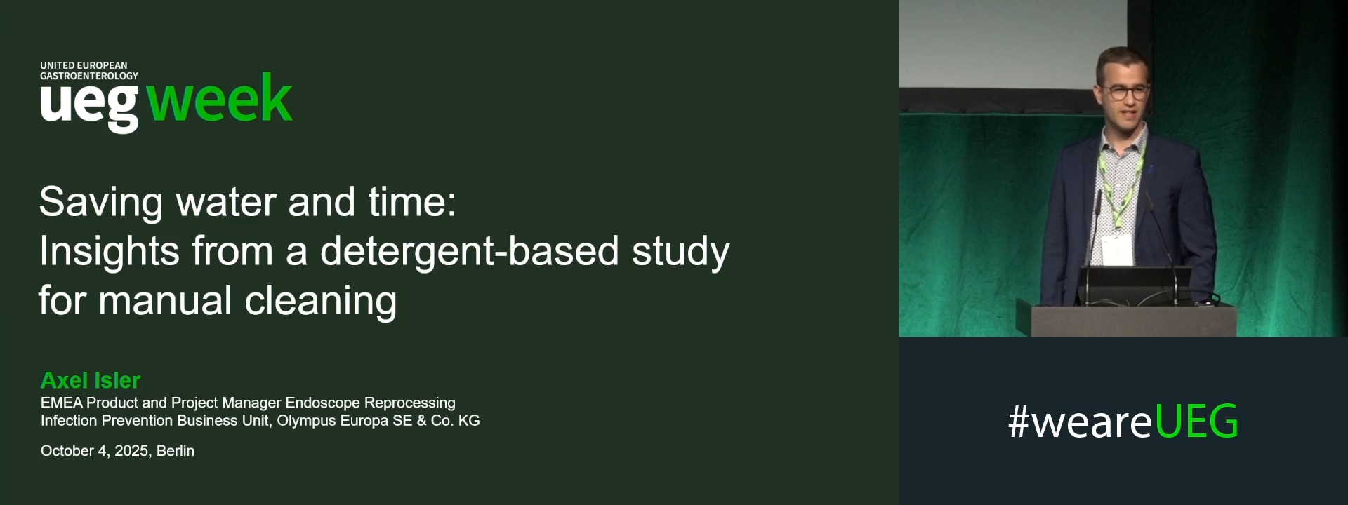
Saving water and time: Insights from a detergent-based study for manual cleaning
1 None, Hamburg, Germany
Event
Topics
Citation
Published
Log in to access this content.
Free for all myUEG account holders. Your access level is set automatically based on your occupation. Medical professionals get full access to all content. If you are a non-medical user, you can only access UEG Week content from congresses you attended.
Not sure what you can access? Learn more about account types.
THE ASSOCIATION OF A POSITIVE FECAL IMMUNOCHEMICAL TEST WITH THE RISK OF GASTROESOPHAGEAL CANCER: A MATCHED COHORT STUDY AND COST-EFFECTIVENESS ANALYSIS
Zohar Levi 1, Naim Abu Freha 2, Maya Aharoni Golan 3, Tom Konikoff 4, Orly Sneh-Arbib 3, Rachel Gingold-Belfer 4, Sapir Eizenstein 5, Doron Boltin 4, Hadar Edelman-Klapper 3, Alexander Vilkin 6, Shiri Kushnir 3, Adi Turgeman 3, Tanya Babitch 3, Moshe Lesnhno 7, Anath Flugelman 8, Elizabeth E Half 8
1 Rabin Medical Center, Petach Tikva, Israel|||Medical and Health Sciences Faculty, Tel Aviv, Israel
2 Soroka University Medical Center, Ben Gurion University of the Negev, Omer, Israel
3 Rabin Medical Center, Petach Tikva, Israel
4 Medical and Health Sciences Faculty, Tel Aviv, Israel
5 Davidoff Cancer Center, Petach Tikva, Israel|||Medical and Health Sciences Faculty, Tel Aviv, Israel
6 Rabin Medical Center, Petah Tikva, Israel
7 Tel Aviv University, Tel Aviv, Israel
8 Rappaport Faculty of Medicine Technion Israeli Institute of Technology, Haifa, Israel
Conference
Topics
Submission format
Citation
Published
Log in to access this content.
Free for all myUEG account holders. Your access level is set automatically based on your occupation. Medical professionals get full access to all content. If you are a non-medical user, you can only access UEG Week content from congresses you attended.
Not sure what you can access? Learn more about account types.
GASTRIC AND DUODENAL CANCER IN LYNCH SYNDROME: INCIDENCE AND YIELD OF UPPER ENDOSCOPY SURVEILLANCE
1 Hospital General Universitario Dr Balmis de Alicante, Alicante, Spain
Conference
Topics
Submission format
Citation
Published
Log in to access this content.
Free for all myUEG account holders. Your access level is set automatically based on your occupation. Medical professionals get full access to all content. If you are a non-medical user, you can only access UEG Week content from congresses you attended.
Not sure what you can access? Learn more about account types.
A UBIQUITOUS AND INTEROPERABLE DEEP LEARNING MODEL FOR AUTOMATIC DETECTION OF PLEOMORPHIC GASTROESOPHAGEAL LESIONS
Miguel Martins 1, Miguel Mascarenhas-Saraiva 1, João Afonso 1, Maria Almeida 1, Tiago Ribeiro 1, Pedro Cardoso 1, Francisco Mendes 1, Joana Mota 1, Joana Frias 1, Catarina Araújo 1, Ana Patrícia Andrade 1, Hélder Cardoso 1, João Ferreira 2, Guilherme Macedo 1
1 São João Univeristy Hospital, Porto, Portugal
2 Faculty of Engineering - University of Porto, Porto, Portugal
Conference
Submission format
Citation
Published
Log in to access this content.
Free for all myUEG account holders. Your access level is set automatically based on your occupation. Medical professionals get full access to all content. If you are a non-medical user, you can only access UEG Week content from congresses you attended.
Not sure what you can access? Learn more about account types.
CLINICAL DECISION TREE FOR OPTIMISING ENDOSCOPIC ASSESSMENT OF SIGNET RING CELL CARCINOMA IN HEREDITARY DIFFUSE GASTRIC CANCER SURVEILLANCE
Lianlian Wu 1, Judith Honing 2, Anjui Wu 3, Sonia S. Kupfer 4, Tanya M. Bisseling 5, Jolanda van Dieren 6, W. Keith Tan 3, Andreas V Hadjinicolaou 7, Florian Markowetz 3, Rebecca Fitzgerald 8, Massimiliano di Pietro 3
1 Cambridge University, Cambridge, United Kingdom
2 University of Cambridge, Rotterdam, Netherlands
3 University of Cambridge, Cambridge, United Kingdom
4 University of Chicago, Chicago, United States
5 Radboud University Medical Centre, Nijmegen, Netherlands
6 Netherlands Cancer Institute, Amsterdam, Netherlands
7 University Of Cambridge, Cambridge, United Kingdom
8 Medical Research Council Hutchison / MRC Research Centre MRC Cancer Unit - MRC Cancer Unit, Medical, Cambridge, United Kingdom
Conference
Topics