Log in to access this content.
Free for all myUEG account holders. Your access level is set automatically based on your occupation. Medical professionals get full access to all content. If you are a non-medical user, you can only access UEG Week content from congresses you attended.
Not sure what you can access? Learn more about account types.
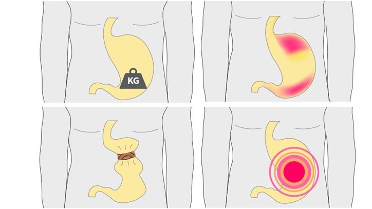
Functional dyspepsia: Diagnosis and treatment
Event
Accreditation status
Duration
Published
Log in to access this content.
Free for all myUEG account holders. Your access level is set automatically based on your occupation. Medical professionals get full access to all content. If you are a non-medical user, you can only access UEG Week content from congresses you attended.
Not sure what you can access? Learn more about account types.
The weird, wonderful , unusual and unexpected in gastroenterology
1 Bradford Teaching Hospitals NHS trust, Leeds, United Kingdom
2 Royal Free Hospital, London, United Kingdom
Published
Log in to access this content.
Free for all myUEG account holders. Your access level is set automatically based on your occupation. Medical professionals get full access to all content. If you are a non-medical user, you can only access UEG Week content from congresses you attended.
Not sure what you can access? Learn more about account types.

IS THIS PRIMARY OR SECONDARY LYMPHOCYTIC OESOPHAGITIS?
1 Klinik Arlesheim, Arlesheim, Switzerland
2 Olten Hospital, Olten, Switzerland
3 Zürich University and Klinik Arlesheim, Arlesheim, Switzerland
Conference
Topics
Submission format
Session
Published
Log in to access this content.
Free for all myUEG account holders. Your access level is set automatically based on your occupation. Medical professionals get full access to all content. If you are a non-medical user, you can only access UEG Week content from congresses you attended.
Not sure what you can access? Learn more about account types.
GASTRIC ENDOSCOPIC SUBMUCOSAL DISSECTION DEFECT RESOLUTION STRATEGIES: THE PATH TO CLOSURE WITH THROUGH-THE-SCOPE HELIX TACK SUTURE SYSTEM
João António Cunha Neves 1, Jéssica Chaves 2, Joana Roseira 1, Mario Dinis-Ribeiro 2, Diogo Libânio 2
1 Unidade Local de Saúde do Algarve, Portimão, Portugal
2 Porto Comprehensive Cancer Center, Porto, Portugal|||MEDCIDS - Department of Community, Medicine, Health Information and Decision, Faculty of Medicine, University of Porto, Porto, Portugal
Conference
Topics
Submission format
Session
Published
Log in to access this content.
Free for all myUEG account holders. Your access level is set automatically based on your occupation. Medical professionals get full access to all content. If you are a non-medical user, you can only access UEG Week content from congresses you attended.
Not sure what you can access? Learn more about account types.
COVID-19 ENCEPHALITIS IN A CIRRHOTIC PATIENT
Sahar Hamza 1, Sabbah Meriam 1, Dorra Trad 1, Houssaina Jlassi 1, Norsaf Bibani 1, Dalila Gargouri 1
1 Habib Thameur Hospital, Tunis, Tunisia
Conference
Submission format
Session
Published
Log in to access this content.
Free for all myUEG account holders. Your access level is set automatically based on your occupation. Medical professionals get full access to all content. If you are a non-medical user, you can only access UEG Week content from congresses you attended.
Not sure what you can access? Learn more about account types.
PRESCRIPTION PATTERN OF PROBIOTICS AS AN ADJUVANT THERAPY FOR HELICOBACTER PYLORI ERADICATION: RESULTS OF THE EUROPEAN REGISTRY ON THE MANAGEMENT OF HELICOBACTER PYLORI INFECTION (HP-EUREG)
Diego Casas Deza 1, Javier Alcedo 1, Miguel Lafuente 2, F. Javier Lopez 2, Ángeles Pérez-Aísa 3, Matteo Pavoni 4, Ilaria Maria Saracino 5, Bojan Tepes 6, Laimas Virginijus Jonaitis 7, Manuel Castro-Fernández 8, Manuel Pabon-Carrasco 8, Alma Keco-Huerga 8, Irina Voynovan 9, Luis Bujanda Fernández de Piérola 10, Alfredo J. Lucendo 11, Natasa Brglez Jurecic 12, Maja Denkovski 12, Perminder S. Phull 13, Luis Ricardo Rodrigo Sáez 14, Angel Lanas 15, Samuel Jesús Martínez-Domínguez 15, Jose Maria Huguet 16, Dmitry S. Bordin 17, Antonio Gasbarrini 18, Juozas Kupcinskas 7, Gülüstan Babayeva 19, Oleksiy Gridnyev 20, Marcis Leja 21, Theodore Rokkas 22, Ricardo Pinto 23, Frode Lerang 24, Doron Boltin 25, Veronika Papp 26, ANTE TONKIC 27, Sinead M. Smith Sinead M. Smith 28, Halis Simsek 29, Marino Venerito 30, Lyudmila Boyanova 31, Vladimir Milivojevic 32, Lumir Kunovsky 33, Tamara Matysiak-Budnik 34, Wojciech Marlicz 35, Michael Doulberis 36, Anna Cano-Catala 37, Luis Hernández Villalba 38, Leticia Moreira Ruiz 39, Olga P. Nyssen 40, Francis Mégraud 41, Colm O'Morain 28, Javier Gisbert 40
1 Miguel Servet University Hospital, Zaragoza, Spain|||Aragon Health Research Institute (IIS Aragon), Zaragoza, Spain
2 University of Zaragoza, Faculty of Sciences, Zaragoza, Spain|||Institute for Biocomputation and Physics of Complex Systems (BIFI). University of Zaragoza, Zaragoza, Spain
3 Agencia Sanitaria Costa del Sol, Marbella, Málaga, Spain
4 IRCCS St. Orsola Polyclinic, University of Bologna, Bologna, Italy|||University of Bologna, Bologna, Italy
5 IRCCS St. Orsola Polyclinic, University of Bologna, Bologna, Italy
6 DC Rogaska, Rogaska Slatina, Slovenia
7 Lithuanian University of Health Sciences, Kaunas, Lithuania
8 Valme University Hospital, Sevilla, Spain
9 A.S. Loginov Moscow Clinical Scientific Center, Moscow, Russian Federation
10 Biodonostia Health Research Institute, San Sebastian, Spain|||Centro de Investigación Biomédica en Red de Enfermedades Hepáticas y Digestivas (CIBERehd), Madrid, Spain|||Universidad del País Vasco (UPV/EHU), San Sebastian, Spain
11 Tomelloso General Hospital, Tomelloso, Ciudad Real, Spain|||Centro de Investigación Biomédica en Red de Enfermedades Hepáticas y Digestivas (CIBERehd), Madrid, Spain|||La Princesa Health Research Institute, Madrid, Spain
12 Interni Oddelek Diagnostic Centre Bled, Bled, Slovenia
13 Aberdeen Royal Infirmary, Aberdeen, United Kingdom
14 University of Oviedo, Oviedo, Spain
15 Hospital Clínico Lozano Blesa, Zaragoza, Spain|||Aragon Health Research Institute (IIS Aragon), Zaragoza, Spain|||Centro de Investigación Biomédica en Red de Enfermedades Hepáticas y Digestivas (CIBERehd), Madrid, Spain
16 University General Hospital of Valencia, Valencia, Spain
17 A.S. Loginov Moscow Clinical Scientific Center, Moscow, Russian Federation|||A.I. Yevdokimov Moscow State University of Medicine and Dentistry, Moscow, Russian Federation|||Tver State Medical University, Tver, Russian Federation
18 Fondazione Policlinico Universitario Agostino Gemelli IRCCS, Roma, Italy
19 Azerbaijan State Advanced Training Institute for Doctors named by A. Aliyev, Baku, Azerbaijan
20 L.T. Malaya Therapy National Institute of the National Academy of Medical Sciences of Ukraine, Kharkiv, Ukraine
21 Gastro, Digestive Diseases Centre, Riga, Latvia|||University of Latvia, Riga, Latvia
22 Henry Dunant Hospital, Athens, Greece
23 Centro Hospitalar do Porto, Porto, Portugal|||Universidade do Porto, Porto, Portugal|||Center for Research in Health Technologies and Information Systems (CINTESIS), Porto, Portugal
24 Østfold Hospital Trust, Grålum, Norway
25 Rabin Medical Center, Petah Tikva, Tel Aviv, Israel|||Tel Aviv University, Tel Aviv, Israel
26 Semmelweis University, Budapest, Hungary
27 University Hospital of Split, Split, Croatia
28 Trinity College Dublin, Dublin, Ireland
29 Hacettepe University, Ankara, Turkey|||HC International Clinic, Ankara, Turkey
30 University Hospital of Magdeburg, Magdeburg, Germany
31 Medical University of Sofia, Sofia, Bulgaria
32 Clinical Center of Serbia, Belgrade, Serbia|||University of Belgrade, Belgrade, Serbia
33 University Hospital Olomouc, Olomuc, Czechia|||Palacky University Olomouc, Olumuc, Czechia|||University Hospital Brno, Brno, Czechia|||Masaryk University, Brno, Czechia
34 University Hospital of Nantes, Nantes, France
35 Pomeranian Medical University in Szczecin, Szczecin, Poland
36 Kantonsspital Aarau, Aarau, Switzerland
37 Althaia Xarxa Assistencial Universitària de Manresa, Manresa, Barcelona, Spain
38 Hospital Santos Reyes, Aranda de Duero, Burgos, Spain
39 Hospital Clínic de Barcelona, Barcelona, Spain|||Centro de Investigación Biomédica en Red de Enfermedades Hepáticas y Digestivas (CIBERehd), Madrid, Spain|||University of Barcelona, Barcelona, Spain
40 La Princesa University Hospital, Madrid, Spain|||La Princesa Health Research Institute (IIS-Princesa), Madrid, Spain|||Centro de Investigación Biomédica en Red de Enfermedades Hepáticas y Digestivas (CIBERehd), Madrid, Spain
41 Université de Bordeaux, Bordeaux, France
Conference
Topics
Submission format
Session
Citation
Published
Log in to access this content.
Free for all myUEG account holders. Your access level is set automatically based on your occupation. Medical professionals get full access to all content. If you are a non-medical user, you can only access UEG Week content from congresses you attended.
Not sure what you can access? Learn more about account types.
INCREASE IN POINT-PREVALENCE AND COSTS OF LIVER CIRRHOSIS IN THE NETHERLANDS – A NATIONWIDE HEALTH CLAIMS DATA ANALYSIS
Koos de Wit 1, Gwen M.C. Masclee 1, Minneke J. Coenraad 2, Frans Cuperus 3, Matthijs Kramer 4, Raoel Maan 5, Robert Bart Takkenberg 1, Marten Alexander Lantinga 1
1 Amsterdam UMC, University of Amsterdam, Amsterdam Gastroenterology Endocrinology Metabolism, Amsterdam, Netherlands
2 Leiden University Medical Centre, Leiden, Netherlands
3 University Medical Center Groningen, Groningen, Netherlands
4 Maastricht University Medical Centre+, Maastricht, Netherlands
5 Erasmus University Medical Center, Rotterdam, Netherlands
Conference
Topics
Submission format
Session