UEG's educational platform for gastroenterology education bringing recordings, courses, guidelines, and more together in one place.
Most popular content
Rate for better recommendations!
Your feedback helps us show you more of what you like and less of what you dislike!
Log in to access this content.
Free for all myUEG account holders. Your access level is set automatically based on your occupation. Medical professionals get full access to all content. If you are a non-medical user, you can only access UEG Week content from congresses you attended.
Not sure what you can access? Learn more about account types.
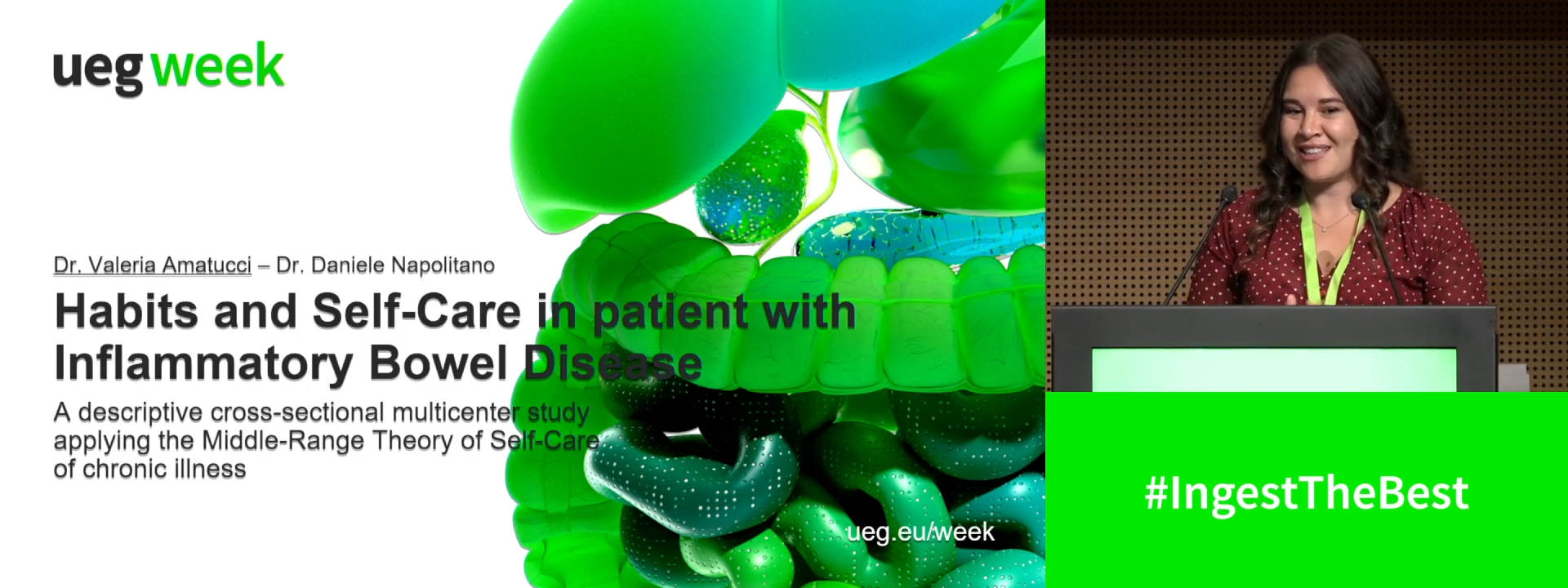
Habits and self-care in patients with Inflammatory Bowel Disease: a descriptive cross-sectional multicenter study applying the Middle Range Theory of self-care of chronic illness
1 None, None, Italy
Event
Topics
Session
Citation
Published
Log in to access this content.
Free for all myUEG account holders. Your access level is set automatically based on your occupation. Medical professionals get full access to all content. If you are a non-medical user, you can only access UEG Week content from congresses you attended.
Not sure what you can access? Learn more about account types.
How to boost your biology? Every day strategies to improve therapies and combinations
1 Charité - Universitätsmedizin Berlin, Berlin, Germany
Event
Citation
Published
Log in to access this content.
Free for all myUEG account holders. Your access level is set automatically based on your occupation. Medical professionals get full access to all content. If you are a non-medical user, you can only access UEG Week content from congresses you attended.
Not sure what you can access? Learn more about account types.
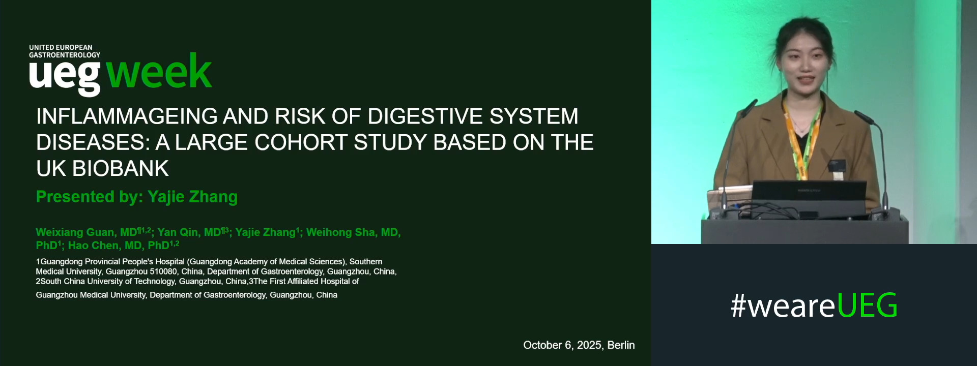
INFLAMMAGEING AND RISK OF DIGESTIVE SYSTEM DISEASES: A LARGE COHORT STUDY BASED ON THE UK BIOBANK
1 Guangdong Provincial People's Hospital (Guangdong Academy of Medical Sciences), Southern Medical University, Guangzhou 510080, China, Guangzhou, China|||South China University of Technology, Guangzhou, China
2 The First Affiliated Hospital of Guangzhou Medical University, Guangzhouchi, China
3 Guangdong Provincial People's Hospital (Guangdong Academy of Medical Sciences), Southern Medical University, Guangzhou 510080, China, Guangzhou, China
Event
Submission format
Session
Citation
Published
Log in to access this content.
Free for all myUEG account holders. Your access level is set automatically based on your occupation. Medical professionals get full access to all content. If you are a non-medical user, you can only access UEG Week content from congresses you attended.
Not sure what you can access? Learn more about account types.
Identifying disease trajectories by AI
1 University Medical Center Schleswig-holstein, Campus Kiel, Kiel, Germany
Event
Topics
Citation
Published
Log in to access this content.
Free for all myUEG account holders. Your access level is set automatically based on your occupation. Medical professionals get full access to all content. If you are a non-medical user, you can only access UEG Week content from congresses you attended.
Not sure what you can access? Learn more about account types.
Fistulising Crohn's disease: Underlying mechanism
1 Icahn School of Medicine at Mount Sinai, New York, United States of America
Event
Topics
Citation
Published
Log in to access this content.
Free for all myUEG account holders. Your access level is set automatically based on your occupation. Medical professionals get full access to all content. If you are a non-medical user, you can only access UEG Week content from congresses you attended.
Not sure what you can access? Learn more about account types.
Optimising success in IBD treatment: Timing surgery and medical treatment
1 Academisch Medisch centrum Dept. of Surgery - Department of Surgery, Academisch Medisch centrum Dept, Amsterdam, Netherlands
2 Royal London Hospital, London, United Kingdom
Event
Citation
Published
Log in to access this content.
Free for all myUEG account holders. Your access level is set automatically based on your occupation. Medical professionals get full access to all content. If you are a non-medical user, you can only access UEG Week content from congresses you attended.
Not sure what you can access? Learn more about account types.
EVALUATION OF SUBCUTANEOUS INFLIXIMAB FOR PERIANAL FISTULA HEALING IN CROHN’S DISEASE: PRELIMINARY RESULTS FROM THE REMSILAP MULTICENTER OBSERVATIONAL COHORT STUDY
Manon Haas 1, Laurent Abramowitz 2, Stephanie Hamonic 3, Amandine Landemaine 3, Guillaume Bouguen 3, Anne Laurain 2, Lucine Vuitton 4, Isabelle Etienney 5, Gauthier Pellet 6, Kristell Coat 3, Ronan Garlantezec 3, Laurent Siproudhis 3, Xavier Treton 1
1 Institut des MICI - Clinique Ambroise Paré Hartmann, Neuilly sur Seine, France
2 CHU Bichat, Paris, France
3 CHU Pontchaillou, Rennes, France
4 Besançon university hospital gastroenterology, Courbevoie, France
5 Diaconesses-Croix St Simon Hospital, Paris, France
6 CHU Bordeaux, Bordeaux, France
Event
Topics