UEG's content platform for gastroenterology education bringing recordings, courses, guidelines, and more together in one place.
Trending on Gutflix
Rate for better recommendations!
Your feedback helps us show you more of what you like and less of what you dislike!
Log in to continue.
This content is part of Gutflix. Log in with your myUEG account, or create one free, to watch it.
Not sure what you can access? Learn more about account types.
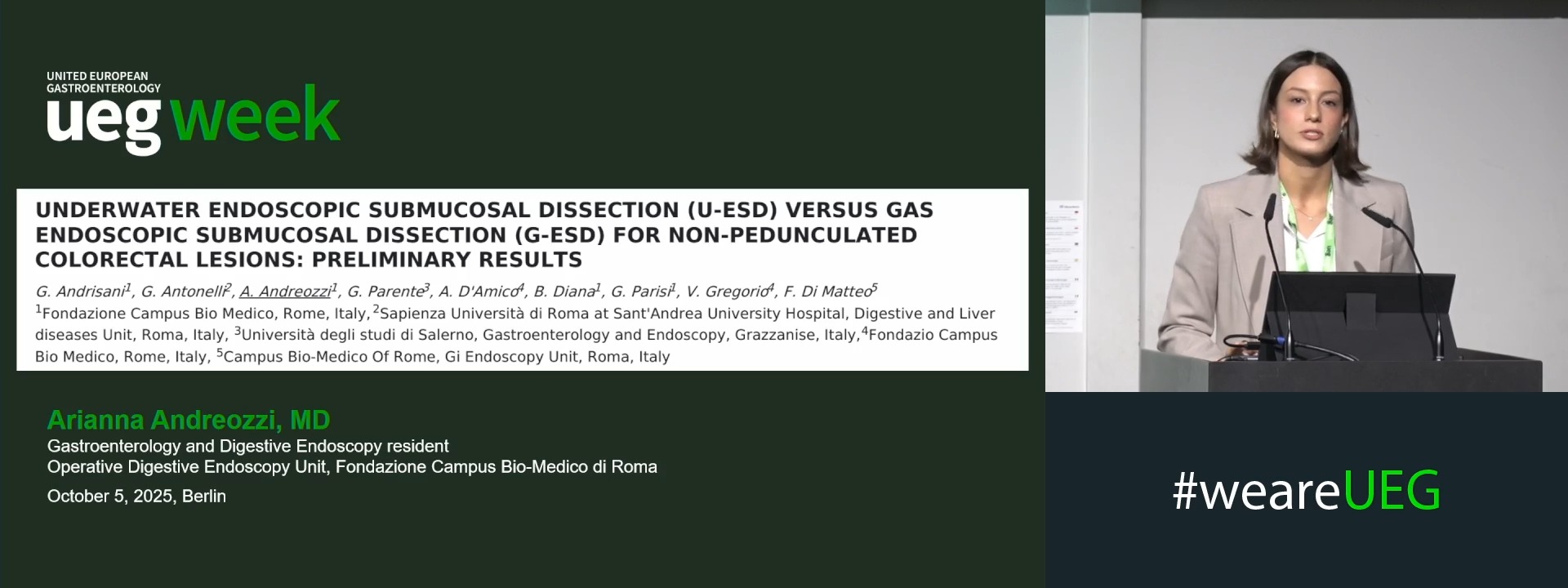
UNDERWATER ENDOSCOPIC SUBMUCOSAL DISSECTION (U-ESD) VERSUS GAS ENDOSCOPIC SUBMUCOSAL DISSECTION (G-ESD) FOR NON-PEDUNCULATED COLORECTAL LESIONS: PRELIMINARY RESULTS
Gianluca Andrisani 1, Giulio Antonelli 2, Arianna Andreozzi 1, Giovanni Parente 3, Andrea D'Amico 4, Benito Diana 1, Giulia Parisi 1, Virginia Gregorio 4, Francesco Di Matteo 5
Affiliations
1 Fondazione Campus Bio Medico, Rome, Italy
2 Sapienza Università di Roma at Sant'Andrea University Hospital, Roma, Italy
3 Università degli Studi di Salerno, Grazzanise, Italy
4 Fondazio Campus Bio Medico, Rome, Italy
5 Campus Bio-Medico of Rome, Roma, Italy
Log in to continue.
This content is part of Gutflix. Log in with your myUEG account, or create one free, to watch it.
Not sure what you can access? Learn more about account types.
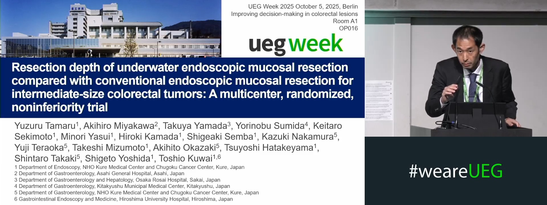
RESECTION DEPTH OF UNDERWATER ENDOSCOPIC MUCOSAL RESECTION COMPARED WITH CONVENTIONAL ENDOSCOPIC MUCOSAL RESECTION FOR INTERMEDIATE-SIZE COLORECTAL TUMORS: A MULTICENTER, RANDOMIZED, NONINFERIORITY TRIAL
Yuzuru Tamaru 1, Akihiro Miyakawa 2, Takuya Yamada 3, Yorinobu Sumida 4, Keitaro Sekimoto 1, Minori Yasui 1, Hiroki Kamada 1, Shigeaki Semba 1, Kazuki Nakamura 1, Yuji Teraoka 1, Takeshi Mizumoto 1, Akihito Okazaki 1, Tsuyoshi Hatakeyama 1, Shintaro Takaki 1, Shigeto Yoshida 1, Toshio Kuwai 5
Affiliations
1 NHO Kure Medical Center and Chugoku Cancer Center, Kure, Japan
2 Asahi General Hospital, Asahi, Japan
3 Osaka Rosai Hospital, Sakai, Japan
4 Kitakyushu Municipal Medical Center, Kitakyushu, Japan
5 Hiroshima University Hospital, Hiroshima, Japan